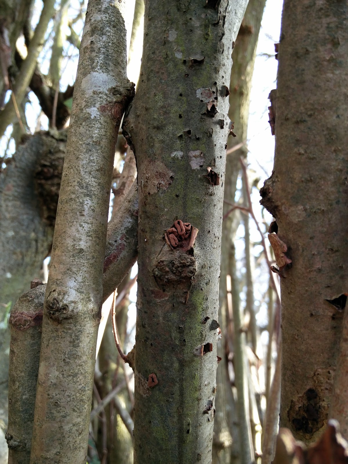
I don't know much about Fungi. What I do know is, they're mostly pretty hard to identify to species. So this is me, trying.
Sunday, 21 May 2017
Off with the Fairies
Found some more mushrooms at Woods Mill today. I think these are Fairy Inkcap Coprinellus disseminatus.
Never seen these before; they look like Glistening Inkcap Coprinellus micaceus but they're much smaller.
For the record
Date: 21/05/2017
Location: Hoe Wood
Grid reference: TQ218136
Saturday, 20 May 2017
Look who's back!
It's been too long since I had some fresh fungus to ID so I took myself off to Woods Mill today with my fungus hunting paraphernalia, determined to find some fruit bodies; hoping the recent rain might have precipitated some action.
Of course I saw all the usual same-old same-old: King Alfred's Cakes Daldinia concentrica, some old Jelly Ear Auricularia auricula-judae, Glue Crust Hymenochaete corrugata and Turkeytail Trametes versicolor.
But nestled in amongst this old Turkeytail Trametes versicolor, I spotted something that looked a bit different.
Algae has turned the Turkeytail Trametes versicolor a beautiful mossy green colour; the pale fruit bodies are something else – Bitter Oysterling Panellus stipticus.
Not super exciting, as I've seen this species before at Woods Mill. But this specimen nicely shows the cracked and 'scurfy' cap surface which is characteristic of this species.
Nearby, I spotted these mushroom-like fruit bodies growing on a well-rotted old branch.
These reminded me of the Bay Polypore Picipes (=Polyporus) badius I found last August, but the fruit bodies are much smaller, less than 4 cm across.
The margin appears quite smooth...
So I think this puts us in the realms of the Winter Polypore Polyporus brumalis; although – according to the books – it's not really the season for it.
Next up, a Trametes species.
This one looks a bit different to the Turkeytail Trametes versicolor that I've got used to seeing everywhere. The brackets are a bit chunkier. Pores are round. And the upper surface feels like chamois leather.
I'm thinking possibly Hairy Bracket Trametes hirsuta, but not 100 % confident.
In Hoe Wood I was very excited to see A MUSHROOM! Growing up through the leaf litter. The stem tapered slightly from a thick base; but I couldn't see any sign of a volva.
It must have just recently sprung up, after the rain, as the gills are still covered by a partial veil.
I didn't want to ruin its reproductive display so I took just a tiny sliver of the cap, to get a look at the gills. They're white, and crowded. And they don't smell of anything in particular.
So, erm, I'm not sure what this is. My first thought was Deathcap Amanita phalloides var. alba. But I can't see any sign of a "large, bag-like white volva". So I'm not convinced it is actually an Amanita. I'll try and get a spore print from my sample and may go back tomorrow for another look.
UPDATE 22/05/2017 – After a misunderstanding with a kerb on Saturday evening, I limped back into Hoe Wood on Sunday to get another look at that white mushroom. It had been knocked over by some woodland creature, since I left it on Saturday. So I thought I'd take it home for a closer look.
After a day in a margarine tub on my desk, the cap has opened to reveal a load of gorgeous chocolate-brown gills. Not white any more! So that's taught me something.
As Ted surmised in the comments below (thanks Ted!) – I can see it's definitely an Agaricus. And it's left a dark brown spore print in my margarine tub.
The key to the Agaricus runs to 12 pages in Funga Nordica. Hmm. We'll have to see how this goes...
*Checks to see if the stem base turns yellow when rubbed* – Nope.
*Checks to see if it smells of bitter almonds* – I don't think so. What do bitter almonds smell like?
*Googles "Schäffer reaction"* – Damn! If only I had some aniline & nitric acid lying around.
*Checks to see if cap goes yellow when bruised* – Hmm... Not really. I'll go to question 11.
*Looks at 'ring'* – It's definitely hanging down.
*Checks to see if flesh in stem top is 'reddening', to some degree* – Um. Don't think so? I'll go to question 26.
Uh oh! I have to look at its spores. *Gets microscope out*
So, the spores look like this:
*Checks to see if the spores have a 'germ pore', or a wall which is 'uniformly thick'* – I can't see any sign of a 'germ pore'. That takes me to question 27.
*Looks at cap again* – Cap is definitely either 'white, whitish or dirty greyish yellowish'. On to question 29...
*Looks to see if the stem is longer than the cap* – Nope, about the same length as cap diameter.
*Has a sniff to see if the smell is unpleasant; metallic* – Don't think so. Just smells kind of mushroom-y to me. This takes me to question 30.
Cripes! I've got to measure the spores now. I haven't done this before, but I did calibrate my new measuring eyepiece the other day, so this could work.
Based on my previous calibration, I think one unit on that scale you can see is about 10 microns. That makes the spore I'm measuring about 7 microns.
*Checks to see if the spores are, on average, < 8 μm long* – Yes! On to question 31.
This takes me to a description of... wait for it... Field Mushroom Agaricus campestris! Well, if it's that, what's it doing growing in the middle of wood?
FURTHER UPDATE 23/05/2017 – Martin Allison emailed. He's not convinced my mushroom's A. campestris. "The ring looks too big and floppy!" He said. So, there we are. The mushroom went in the bin last night, so it's identity must remain forever a mystery.
Last up, my old friend Chicken of the Woods Laetiporus sulphureus, growing on the same log where we found it last year.
What a beauty!
I grew quite attached to this fungus during its fruiting season last year. I watched it develop from plump, peachy pillows into bright frilly brackets. It seemed quite beautiful to me even as it slipped into senescence; until it finally fell to earth on the day of the referendum result. True story.
For the record
Date: 20/05/2017
Location: Hoe Wood
Grid reference: TQ217136
D. concentrica, P. stipticus, P. brumalis & L. sulphureus entered into FRDBI database 06/09/2018
Of course I saw all the usual same-old same-old: King Alfred's Cakes Daldinia concentrica, some old Jelly Ear Auricularia auricula-judae, Glue Crust Hymenochaete corrugata and Turkeytail Trametes versicolor.
But nestled in amongst this old Turkeytail Trametes versicolor, I spotted something that looked a bit different.
Algae has turned the Turkeytail Trametes versicolor a beautiful mossy green colour; the pale fruit bodies are something else – Bitter Oysterling Panellus stipticus.
Not super exciting, as I've seen this species before at Woods Mill. But this specimen nicely shows the cracked and 'scurfy' cap surface which is characteristic of this species.
Nearby, I spotted these mushroom-like fruit bodies growing on a well-rotted old branch.
![]() |
| Look! No gills. |
The margin appears quite smooth...
So I think this puts us in the realms of the Winter Polypore Polyporus brumalis; although – according to the books – it's not really the season for it.
Next up, a Trametes species.
I'm thinking possibly Hairy Bracket Trametes hirsuta, but not 100 % confident.
In Hoe Wood I was very excited to see A MUSHROOM! Growing up through the leaf litter. The stem tapered slightly from a thick base; but I couldn't see any sign of a volva.
It must have just recently sprung up, after the rain, as the gills are still covered by a partial veil.
I didn't want to ruin its reproductive display so I took just a tiny sliver of the cap, to get a look at the gills. They're white, and crowded. And they don't smell of anything in particular.
So, erm, I'm not sure what this is. My first thought was Deathcap Amanita phalloides var. alba. But I can't see any sign of a "large, bag-like white volva". So I'm not convinced it is actually an Amanita. I'll try and get a spore print from my sample and may go back tomorrow for another look.
UPDATE 22/05/2017 – After a misunderstanding with a kerb on Saturday evening, I limped back into Hoe Wood on Sunday to get another look at that white mushroom. It had been knocked over by some woodland creature, since I left it on Saturday. So I thought I'd take it home for a closer look.
After a day in a margarine tub on my desk, the cap has opened to reveal a load of gorgeous chocolate-brown gills. Not white any more! So that's taught me something.
As Ted surmised in the comments below (thanks Ted!) – I can see it's definitely an Agaricus. And it's left a dark brown spore print in my margarine tub.
The key to the Agaricus runs to 12 pages in Funga Nordica. Hmm. We'll have to see how this goes...
*Checks to see if the stem base turns yellow when rubbed* – Nope.
*Checks to see if it smells of bitter almonds* – I don't think so. What do bitter almonds smell like?
*Googles "Schäffer reaction"* – Damn! If only I had some aniline & nitric acid lying around.
*Checks to see if cap goes yellow when bruised* – Hmm... Not really. I'll go to question 11.
*Looks at 'ring'* – It's definitely hanging down.
*Checks to see if flesh in stem top is 'reddening', to some degree* – Um. Don't think so? I'll go to question 26.
Uh oh! I have to look at its spores. *Gets microscope out*
So, the spores look like this:
*Checks to see if the spores have a 'germ pore', or a wall which is 'uniformly thick'* – I can't see any sign of a 'germ pore'. That takes me to question 27.
*Looks at cap again* – Cap is definitely either 'white, whitish or dirty greyish yellowish'. On to question 29...
*Looks to see if the stem is longer than the cap* – Nope, about the same length as cap diameter.
*Has a sniff to see if the smell is unpleasant; metallic* – Don't think so. Just smells kind of mushroom-y to me. This takes me to question 30.
Cripes! I've got to measure the spores now. I haven't done this before, but I did calibrate my new measuring eyepiece the other day, so this could work.
Based on my previous calibration, I think one unit on that scale you can see is about 10 microns. That makes the spore I'm measuring about 7 microns.
*Checks to see if the spores are, on average, < 8 μm long* – Yes! On to question 31.
This takes me to a description of... wait for it... Field Mushroom Agaricus campestris! Well, if it's that, what's it doing growing in the middle of wood?
FURTHER UPDATE 23/05/2017 – Martin Allison emailed. He's not convinced my mushroom's A. campestris. "The ring looks too big and floppy!" He said. So, there we are. The mushroom went in the bin last night, so it's identity must remain forever a mystery.
Last up, my old friend Chicken of the Woods Laetiporus sulphureus, growing on the same log where we found it last year.
What a beauty!
I grew quite attached to this fungus during its fruiting season last year. I watched it develop from plump, peachy pillows into bright frilly brackets. It seemed quite beautiful to me even as it slipped into senescence; until it finally fell to earth on the day of the referendum result. True story.
![]() |
| Chicken of the Woods Laetiporus sulphureus at Woods Mill, 2016. IMAGE | Clare Blencowe. |
For the record
Date: 20/05/2017
Location: Hoe Wood
Grid reference: TQ217136
D. concentrica, P. stipticus, P. brumalis & L. sulphureus entered into FRDBI database 06/09/2018
Monday, 20 March 2017
Sorting Elfcups
I had the pleasure of joining Sussex Fungus Group once again on Saturday for a spring foray around Sussex Wildlife Trust's Woods Mill nature reserve. It was a treat for me to go round this site, which I visit all the time, with an experienced mycologist: Nick Aplin. A valuable opportunity to find out what I've been getting wrong!
As I've probably mentioned before on this blog, it's difficult to know what you don't know when you're getting into mycology. And this weekend turned up a classic example.
One of my colleagues, Renzo Spano, first spotted these bright red fruiting bodies on 1 March and they were still putting on a good show, next to the footpath which skirts the south-western side of the lake, when we visited on Saturday.
I identified them immediately as Scarlet Elfcup Sarcoscypha austriaca, a distinctive species featured in the Collins Complete (photographic) Guide which I'd seen popping up all over social media during the preceding couple of weeks.
What the Collins Complete (photographic) Guide didn't tell me – what I would have found out if I'd bothered to look in the slightly more weighty Collins (illustrated) Fungi Guide – is that there's another species which looks almost exactly the same as Scarlet Elfcup S. austriaca: the Ruby Elfcup S. coccinea.
Nick explained that the two can be separated fairly easily with microscopy, so I thought I'd have a go. If you google Sarcoscypha you can find some nice accessible blogs which tell you what to look for, like this one on first-nature.com and this one by Michael Kuo. And Liz Holden tells the story of these two species on the Scottish Fungi website.
Armed with this information, I mounted a thin section of the fruit body in water and placed it under the microscope.
Here's an image of one of the 'asci', i.e. one of the long thin sacs in which the 'ascospores' are formed. There are eight ascospores in this ascus. I think both S. austriaca and S. coccinea can have eight spores per ascus, so this doesn't really get me anywhere but it's quite pretty so I thought I'd stick it in. The red stuff you can see are 'paraphyses' - thin threads which give the fruit bodies their distinctive colour.
Nick had mentioned that a key feature of S. austriaca is that its ascospores sometimes appear depressed at the end. I think this might be a depressed ascospore...
Also, the ascospores of S. austriaca sometimes produce 'conidia' (asexual spores) on the ends. I might have found some of these. Not sure.
Finally, Liz Holden suggests that an easy way to tell these two species apart is by looking at the hairs on the outer surface of the cup. She explains that "S. austriaca has hairs that are almost corkscrewed in appearance whereas in S. coccinea they are straight or gently curved at most."
This is what I observed on the outer surface of the cup. Quite screwy?
With all this additional information, I'm inclined to think my first guess was – by chance – correct, and this is Scarlet Elfcup S. austriaca. But would be good to get that confirmed by someone who knows what they're talking about...
UPDATE 22/03/2017 - have received confirmation from the Sussex County Recorder for Ascomycetes, Nick Aplin, that this "100 % S. austriaca". Go me!
For the record
Date (of collection): 18 March 2017
Location: Woods Mill
Grid reference: TQ21821365
Record previously entered into FRDBI on 1 March as S. austriaca
As I've probably mentioned before on this blog, it's difficult to know what you don't know when you're getting into mycology. And this weekend turned up a classic example.
One of my colleagues, Renzo Spano, first spotted these bright red fruiting bodies on 1 March and they were still putting on a good show, next to the footpath which skirts the south-western side of the lake, when we visited on Saturday.
![]() |
| Bright red cup fungi, but what species? |
What the Collins Complete (photographic) Guide didn't tell me – what I would have found out if I'd bothered to look in the slightly more weighty Collins (illustrated) Fungi Guide – is that there's another species which looks almost exactly the same as Scarlet Elfcup S. austriaca: the Ruby Elfcup S. coccinea.
Nick explained that the two can be separated fairly easily with microscopy, so I thought I'd have a go. If you google Sarcoscypha you can find some nice accessible blogs which tell you what to look for, like this one on first-nature.com and this one by Michael Kuo. And Liz Holden tells the story of these two species on the Scottish Fungi website.
Armed with this information, I mounted a thin section of the fruit body in water and placed it under the microscope.
Here's an image of one of the 'asci', i.e. one of the long thin sacs in which the 'ascospores' are formed. There are eight ascospores in this ascus. I think both S. austriaca and S. coccinea can have eight spores per ascus, so this doesn't really get me anywhere but it's quite pretty so I thought I'd stick it in. The red stuff you can see are 'paraphyses' - thin threads which give the fruit bodies their distinctive colour.
Nick had mentioned that a key feature of S. austriaca is that its ascospores sometimes appear depressed at the end. I think this might be a depressed ascospore...
Also, the ascospores of S. austriaca sometimes produce 'conidia' (asexual spores) on the ends. I might have found some of these. Not sure.
This is what I observed on the outer surface of the cup. Quite screwy?
With all this additional information, I'm inclined to think my first guess was – by chance – correct, and this is Scarlet Elfcup S. austriaca. But would be good to get that confirmed by someone who knows what they're talking about...
UPDATE 22/03/2017 - have received confirmation from the Sussex County Recorder for Ascomycetes, Nick Aplin, that this "100 % S. austriaca". Go me!
For the record
Date (of collection): 18 March 2017
Location: Woods Mill
Grid reference: TQ21821365
Record previously entered into FRDBI on 1 March as S. austriaca
Wednesday, 15 February 2017
Peniophora sp.?
On my trip to Horton Wood on Monday, searching for Spring Hazelcup Encoelia furfuracea, I came across this dried-out, cracked-up resupinate fungus.
I thought it was growing on Hazel, but my winter tree-identification still leaves a lot to be desired.
One of the things I've found difficult, in getting into fungus recording, is it's very hard to know what you don't know.
Starting out with the Collins Complete (photographic) Guide, I'd have been inclined to call this something like Peniophora cinerea (one of the five Peniophora species that gets a mention in the Collins book)...
However, having recently purchased The Resupinates of Hampshire (2017), I now realise there are 17 different Peniophora species featured in that guide; many of which look, well, rather similar.
So I think I'll leave this here, until I've learnt a bit more about resupinates.
For the record
Date: 13/02/2017
Location: Horton Wood
Grid reference: TQ208127
I thought it was growing on Hazel, but my winter tree-identification still leaves a lot to be desired.
One of the things I've found difficult, in getting into fungus recording, is it's very hard to know what you don't know.
Starting out with the Collins Complete (photographic) Guide, I'd have been inclined to call this something like Peniophora cinerea (one of the five Peniophora species that gets a mention in the Collins book)...
However, having recently purchased The Resupinates of Hampshire (2017), I now realise there are 17 different Peniophora species featured in that guide; many of which look, well, rather similar.
So I think I'll leave this here, until I've learnt a bit more about resupinates.
For the record
Date: 13/02/2017
Location: Horton Wood
Grid reference: TQ208127
This is what Spring Hazelcup habitat looks like
Following the return of Spring Hazelcup Encoelia furfuracea to Hoe Wood earlier this week, as I mentioned here, I thought I'd get out and check the other sites round the village where I found fruiting bodies last year.
Scanning the overgrown hazels which border one of the footpaths to the west of the village, I eventually struck lucky with this one.
Spring Hazelcup E. furfuracea fruit bodies were just emerging from one of the vertical branches.
So that's fruit bodies re-found at two out of three sites where this species occurred last year.
I went on to check the straggly hazel tree where I first found Spring Hazelcup E. furfuracea, in January 2015, but found no sign of it there.
I did, however, find another little outcrop of Spring Hazelcup E. furfuracea growing on one of the old coppice hazels in Horton Wood, here:
Not bad for an afternoon's work.
For the record
Date: 13/02/2017
Location: New Hall public footpath, Small Dole & Horton Wood, Small Dole
Grid reference: TQ20851311 & TQ20801272
Entered into FRDBI: 15/02/2017
Scanning the overgrown hazels which border one of the footpaths to the west of the village, I eventually struck lucky with this one.
![]() |
| Spring Hazelcup E. furfuracea habitat, New Hall footpath, Small Dole. |
Spring Hazelcup E. furfuracea fruit bodies were just emerging from one of the vertical branches.
So that's fruit bodies re-found at two out of three sites where this species occurred last year.
I went on to check the straggly hazel tree where I first found Spring Hazelcup E. furfuracea, in January 2015, but found no sign of it there.
I did, however, find another little outcrop of Spring Hazelcup E. furfuracea growing on one of the old coppice hazels in Horton Wood, here:
![]() |
| Spring Hazelcup E. furfuracea habitat, Horton Wood, Small Dole |
Not bad for an afternoon's work.
For the record
Date: 13/02/2017
Location: New Hall public footpath, Small Dole & Horton Wood, Small Dole
Grid reference: TQ20851311 & TQ20801272
Entered into FRDBI: 15/02/2017
Sunday, 12 February 2017
Guess who's back!
I've been periodically returning to the sites where I found Spring Hazelcup Encoelia furfuracea last year to see if I could find any further sign of this charismatic little brown fungus. The fruit bodies disappeared entirely after fruiting and I wasn't sure if they'd come again; 2016 seemed to be a particularly good year for them with my finds around Small Dole and Nick Aplin's find at Tilgate Park, Crawley.
I've seen no sign of E. furfuracea since early spring 2016. Until today! On a quick walk through Hoe Wood with Vivien Hodge, I found these – emerging from the same dead Hazel branch where I found them last year. It's back!
I found it here on 20 February last year, so they've re-appeared at a very similar time of year.
This can only mean one thing: time to resume the Small Dole Spring Hazelcup Survey! So far I've found this species at three locations around the village; it seems to favour standing dead Hazel / neglected Hazel coppice. Am keen to see if it crops up anywhere else.
Worth keeping an eye out for it at other sites in Sussex as I believe it's only been recorded in a handful of places in the county.
For the record
Date: 12/02/2017
Location: Hoe Wood [private site]
Grid reference: TQ21821350
Entered into FRDBI: 12/02/2017
I've seen no sign of E. furfuracea since early spring 2016. Until today! On a quick walk through Hoe Wood with Vivien Hodge, I found these – emerging from the same dead Hazel branch where I found them last year. It's back!
![]() |
| Spring Hazelcup Encoelia furfuracea in Hoe Wood, 12/02/2017 |
This can only mean one thing: time to resume the Small Dole Spring Hazelcup Survey! So far I've found this species at three locations around the village; it seems to favour standing dead Hazel / neglected Hazel coppice. Am keen to see if it crops up anywhere else.
Worth keeping an eye out for it at other sites in Sussex as I believe it's only been recorded in a handful of places in the county.
For the record
Date: 12/02/2017
Location: Hoe Wood [private site]
Grid reference: TQ21821350
Entered into FRDBI: 12/02/2017
Saturday, 21 January 2017
The old oak tree
It's been a frosty week in Small Dole and the ground was frozen solid as I headed off for a jaunt this afternoon. There was not a single mushroom to be seen.
On the fallen bough of this oak tree, I caught a climpse of an old crust fungus, so I thought I'd take a closer look.
It seemed like some considerable time has passed since since the break, as much of the upper section had lost its bark. Swathes of orangey-brown crust fungus covered one of the limbs, in tiered rows with ruffled edges – looking well past their best: one of the Crusts, a Stereum or Hymenochaete species, but I wasn't sure how to determine which it is, especially with it being so old.
On one of the higher limbs, where the bark had fallen away, I spotted a fresh streak of crust fungus and thought this might give me a better chance at an identification.
The hairy upper surface and smooth, orange-coloured underside makes me think this is Hairy Curtain Crust Stereum hirsutum.
Several of the limbs, where the bark was still attached, were covered in a smattering of these.
Looking in the Collins Complete (photographic) Guide, it seems likely these are Brown Cup Rutstroemia firma. They're attached to the substrate by a shortish stem (yup – checked that) and grow gregariously on dead branches of deciduous trees, especially oaks and Hazel.
I liked them.
Having now totted up three species on this old oak bough, on this still-frosty January day, I thought I'd see how many more I could get.
There was this knobbly Phellinus type thing, which I'm not even going to attempt to identify...
... which may or may not be the same species as this.
Also quite a lot of this:
It was interesting to see the difference in growing habit between the upper and lower surface of the bough: they definitely know which way is up.
I'm going to take a punt on that one being Split Porecrust Schizopora paradoxa.
Moving from white to black, I'm pretty sure the black, jelly-like flaps attached to this oak twig are Witches' Butter Exidia glandulosa.
And this black mat was very striking – growing over a mossy substrate.
Looking at it under the stereomicroscope, there's very little 'structure' to it. It's just a mass of jet-black threads.
I think these may be the hyphae of Chaetosphaerella phaeostroma, which I've had in Horton Wood before.
Pretty happy with that little haul.
For the record
Date: 21/01/2017
Location: Horton Wood, Small Dole
Grid reference: TQ2078612801 [grid reference is for the fallen oak bough]
Entered into FRDBI: 12/02/2017
On the fallen bough of this oak tree, I caught a climpse of an old crust fungus, so I thought I'd take a closer look.
It seemed like some considerable time has passed since since the break, as much of the upper section had lost its bark. Swathes of orangey-brown crust fungus covered one of the limbs, in tiered rows with ruffled edges – looking well past their best: one of the Crusts, a Stereum or Hymenochaete species, but I wasn't sure how to determine which it is, especially with it being so old.
On one of the higher limbs, where the bark had fallen away, I spotted a fresh streak of crust fungus and thought this might give me a better chance at an identification.
The hairy upper surface and smooth, orange-coloured underside makes me think this is Hairy Curtain Crust Stereum hirsutum.
Several of the limbs, where the bark was still attached, were covered in a smattering of these.
Looking in the Collins Complete (photographic) Guide, it seems likely these are Brown Cup Rutstroemia firma. They're attached to the substrate by a shortish stem (yup – checked that) and grow gregariously on dead branches of deciduous trees, especially oaks and Hazel.
I liked them.
Having now totted up three species on this old oak bough, on this still-frosty January day, I thought I'd see how many more I could get.
There was this knobbly Phellinus type thing, which I'm not even going to attempt to identify...
... which may or may not be the same species as this.
Also quite a lot of this:
It was interesting to see the difference in growing habit between the upper and lower surface of the bough: they definitely know which way is up.
I'm going to take a punt on that one being Split Porecrust Schizopora paradoxa.
Moving from white to black, I'm pretty sure the black, jelly-like flaps attached to this oak twig are Witches' Butter Exidia glandulosa.
And this black mat was very striking – growing over a mossy substrate.
Looking at it under the stereomicroscope, there's very little 'structure' to it. It's just a mass of jet-black threads.
Pretty happy with that little haul.
For the record
Date: 21/01/2017
Location: Horton Wood, Small Dole
Grid reference: TQ2078612801 [grid reference is for the fallen oak bough]
Entered into FRDBI: 12/02/2017
Subscribe to:
Posts (Atom)