We finished work early on Tuesday to fit in a fungi walk before dinner and drove out, past Beggars Bush, to Spithandle Lane.
Sprouting up from the edge of Spithandle Lane we spotted this. Ridiculous!
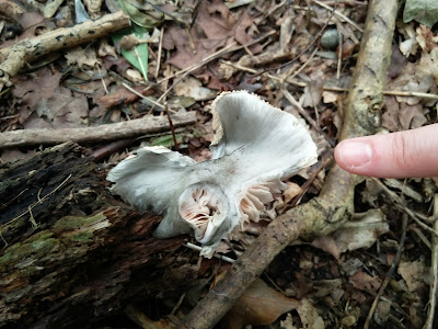
And nearby, another fruit body which had spread its gills and now stood proud, above the woodland floor.
These look like the
Parasol Macrolepiota procera; the brown scales on the stem, giving it a snakeskin-like appearance, are a key characteristic.
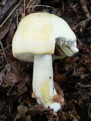
Nearby, this mushroom was also hard to miss.
Underneath the cap, a partial veil entirely covered its gills.
It looked so utterly perfect, I was reluctant to start poking at it to discover more of its features. Perhaps it's a beautiful white
Death Cap Amanita phalloides var.
alba. I should have at least sniffed it, to see if I could detect its sweet deathly scent.
Our route took us south, through mixed woodland, and I was soon overwhelmed by the number of fungi fruiting here.
Little Brown Jobs, Russulas and most of the samey-looking Boletes were brazenly ignored; while I scanned the woodland floor for fruit bodies I might have a cat-in-hell's chance of identifying.
Aha! One I think I know:
Oak Curtain Crust Hymenochaete rubiginosa.
And one I definitely don't know. But which was too pretty to pass by.
I noticed fragments of... veil (?) hanging from the edges of the rounded white cap.
Not sure what genus I'm in with this one:
Psathyrella? Perhaps a young
Pale Brittlestem Psathyrella candolleana. I moved on.
I found this mushroom growing amongst the leaflitter:
Amethyst Deceiver Laccaria amethystina.
I wondered if this emerging mushroom might be a
Tawny Grisette Amanita fulva. A volva 'flushed with cap colour' is characteristic of this species.
On a rotting log I spotted these flat fruit bodies.
Underneath: pores.
It looks like there are also the remains here of a dead and blackened stipe, perhaps of a previous generation of these fruit bodies. I wonder if these are young specimens of the
Blackfoot Polypore Polyporus leptocephalus.
On the same rotting log: YELLOW BLOBS!
I do love a good blob. I felt this must be slime mould plasmodium, for sure, so I took a sample home to see what it would do.
By the time I got home from work the next day, it looked like this:
 |
| Photographed down stereomicroscope. |
Reddish-brown tubes on stalks. Amazing! Wish I'd seen this in progress.
I believe that makes this one of the
Stemonitis species of slime mould. There are a couple which Bruce Ing, in his book 'The Myxomycetes of Britain and Ireland', describes as having yellow plasmodium and occuring on rotten wood:
- Stemonitis axifera
- Stemonitis flavogenita
They're quite similar!
S. flavogenita is reported to have a 'zigzag' at the top of the column ('columella') which supports the spore-bearing structure ('sporangium'). I couldn't see anything like that. But other features, such as the blunt sporangia, do seem to point to
S. flavogenita. I think I'll have to pass on this one.
UPDATE 12/08/2017: I've received some very helpful advice from Malcolm Storey, over on the British Slime Moulds (Myxogastria) Facebook Page, on how to check for the presence of that 'zigzag' which is indicative of S. flavogenita. He advises blowing all the spores off and then getting one or two sporangia mounted in water (with a drop of detergent), under a compound microscope. You might need to keep tapping and rinsing it to wash all the spores off.
If it's there, the kink should look like this:
 |
| Copyright © Malcolm Storey, 2002, www.bioimages.org.uk. Some rights reserved. |
I had a go at doing this but it seems my specimen is unfortunately now too mangled to determine if it is or isn't kinked.
Further on, where the path took us along the edge of a pasture, with a few oaks growing along the edges, I spotted what I presume is another
Inocybe.
A quick up-stipe shot revealed pale-edged gills, like I'd observed on that
Inocybe in Horton Wood the other day.
As I got stuck trying to identify the last
Inocybe I collected, I thought I wouldn't force myself to attempt another, just yet. So I left this mushroom as I found it.
Michael spotted the large cap of an old
Amanita collapsed at the bottom of an oak tree. I think this may be a
Blusher Amanita rubescens, but I wouldn't like to say for sure without seeing more of it.
Entering an area of Birch woodland, we were suddenly surrounded by
earthballs Scleroderma sp. I'm not 100 % confident identifying earthballs, but I think those coarse scales on the surface might make these
Common Earthball Scleroderma citrinum.
A little further on, I noticed this streak of mushrooms which stretched for about a metre to that tree in the background, and then continue for another metre or two on the other side.
I'd decided to ignore Little Brown Jobs but these mushrooms had such a distinctive growing habit, I thought it would be worth stopping for a look.
All along the line, the mushrooms were growing in tightly packed clusters; the long, downy stems actually fused together, up to perhaps a quarter of their length:
Having flicked through the my field guides, I'm going to hazard a guess at
Clustered Toughshank Gymnopus (=
Collybia)
confluens; the field characteristics match the descriptions very well. If I'm right, they should provide a white spore print
– I'll see if I can get one (although the mushrooms are now rather dried out).
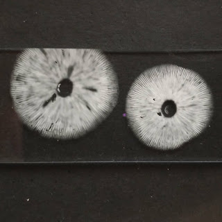
UPDATE 14/08/2017: Managed to get a spore print from a couple of those clustered mushrooms. Definitely white!
Moving on, into an area of more mixed woodland, a definite
Tawny Grisette Amanita fulva.
It was growing next to these mousy-brown mushroom, which had me stumped at first.
But then I saw that they were growing from a chunk of rotting dead wood.
At this point, I decided they were
Deer Shield Pluteus cervinus. However, as I am writing this, I notice that the gills in the photograph above are not 'crowded', as you'd expect in
P. cervinus. So perhaps I got this one wrong.
We then came across these gorgeous
Amanitas growing at the edge of a plantation woodland, with cypresses
.
I think this is another example of
The Blusher Amanita rubescens.
There were quite a few around. In this specimen, the flawless partial veil still clung to the edges of the cap.
What a beauty.
Moving quickly on, because by this time dinner was calling...
Yellow Stagshorn Calocera viscosa. Looking GREAT.
Chanterelle Cantharellus cibarius.
These orange mushrooms were growing out of a pile of rotting logs.
Orange gills makes these one of the Rustgills
gymnopilus sp. I think.
I'm going to put my money on
Common Rustgill Gymnopilus penetrans.
I saw some more of these
– a common sight on my forays this summer:
Spindleshank Gymnopus fusipes.
And finally, I should mention the couple of boletes which I decided
not to ignore.
There was this one, which I got very excited about.
Which ended up being
Chestnut Bolete Gyroporus castaneus, as I established in
an earlier blog post.
Then there was this dark, suede-topped beauty growing at the edge of a conifer plantation, which my photos really don't do justice to.
So, what can we see here.
- Cap surface: dry and velvety, almost suede-like.
- Stem: smooth; base colour is yellow, with red dots and longditudinal striations.
- Tubes: when split tear in half lengthways
This takes us to the
Xerocomoid boletes in Geoffrey Kibby's 'British Boletes' (2016).
I
think that key takes me to
Red Cracking Bolete Xerocomellus chrysenteron. The olive-brown spore print would support that identification.
I had the impression that the fruit body I collected was a young specimen (which got knocked around a bit before I had a chance to take the photos above); which might explain why it doesn't show any cracking.
THE END! Ah, I thought I'd been fairly selective in which fungi I stopped to investigate on our Tuesday night foray. It's still taken me most of the week to puzzle my way through them. Now, what will this weekend bring?
For the record
Date: 08/08/2017
Location: Near Spithandle Lane, Ashurst, West Sussex
Grid reference: TQ11S
Records entered into FRDBI 06/09/2018


