Sunday 3 December 2017

Making Connections

I've had quite a bit going on recently, so it was a couple of weeks ago now that I spent Thursday and Friday in Cardiff at the National Biodiversity Network (NBN) conference before heading off to the British Mycological Society's Open Autumn Meeting at the Jodrell Laboratory, Kew, on Saturday 18 November.

I'm not used to this level of gadding-about and hob-nobbing so by the time I got home on Saturday evening my brain was buzzing. I normally try and keep some separation between my work as a local environmental record centre manager at Sussex Biodiversity Record Centre and the fungus recording which I do as a hobby; so the mycology doesn't start to feel like 'work'. But on this occasion they all got tangled up together.

This blog is an attempt at turning that tangle into something vaguely coherent.

I noticed a few weeks back that the British Mycological Society (BMS) had become a member of the National Biodiversity Network (NBN).
This struck me as a very positive development, as the NBN includes lots of different organisations, schemes and societies with an interest in biological data. And I'm all for working together.

One theme that often comes up in NBN conference talks is how to get more and better biological data; and make it accessible so it can be used. On this topic, Sue Townsend gave a short update on the Field Studies Council's exciting new BioLinks project which has been awarded Heritage Lottery funding to support communities of volunteers in developing biological recording skills. The project will focus particularly on difficult and under-represented taxa and helping people to access the mentoring and resources that are needed to build expertise and confidence in recording these groups. 

During the consultation phase for the BioLinks project (which was managed very effectively by Keiron Derek Brown) I did my best to make a play for fungi being one of the 'difficult and under-represented taxa' that the project might focus on. But it seems the invertebrates got the most votes in the end. Still, I'm sure many of the BioLinks principles and ideas are transferrable so I'd urge folk interested in this kind of thing to take a look at the consultation report and development plan for training provision which the folk at FSC have been kind enough to share.

Listening to the talks at the BMS Open Autumn Meeting on the Saturday, it struck me that the challenges with recording fungi are on a different scale to many other taxon groups. Not least because there is still so much work to do on taxonomy.

I'm not the first person to think this: Caroline Hobart, in her talk, referenced a report from the House of Lords Science and Technology Committee which concluded that, "the state of systematics and taxonomy in the UK, both in terms of the professional taxonomic community and volunteers, is unsatisfactory – in some areas, such as mycology, to the point of crisis." Gosh.

After being rather scared off the Cortinarius last year (which I talked about here), I was very interested to hear Dr. Jorinde Nuytinck explain the work she's been doing on the European Milkcaps (Lactarius and Lactifluus species): looking at the extent to which species grouped according to their morphological characters ('morpho-species') match up with phylogenetic species concepts (i.e. an understanding of whether species are descended from a common ancestor, based on DNA analysis).

As is the case with most fungus talks I go to these days, this resulted in me discovering that the one Milkcap I thought I knew, the Ugly Milkcap Lactarius turpis is not what I thought it was: the recommended name is now Lactarius necator. And I think Dr. Nuytinck said something about it being cryptic, along with Lactarius sordidus (?), i.e. these two species are, according to our current understanding, morphologically identical but phylogenetically distinct. Like a 'brother from another mother'?

DNA barcoding sounds like it's not at all straight forward and can lead you up the garden path if you're not careful (and there was me imagining it to be some kind of magic answer machine). So it was good to hear Dr. Nuytinck explain the different techniques she's used to ensure she is drawing the correct inferences about species' relatedness and phylogeny.

Overall the work of earlier taxonomists seems to have stood up remarkably well to scrutiny as Dr. Nuytinck explained that 75 % of the previously-named European Milkcap species are phylogenetic species. I guess there's still some sorting out to do on the remaining 25 %.

Later on that morning Prof. Henry Beker gave us an update on the Hebeloma. And I think the short summary of that talk would be... these are DONE! After decades of research, Prof. Beker and his colleagues have now sorted out the phylogenetic species of Hebeloma and, through pain-staking observation and databasing, found morphological features which can be used to separate the various species. This work has been published in the latest issue of Field Mycology which landed on my doormat a few days ago, in an article on 'Hebeloma in the United Kingdom', and also in a new volume of Fungi Europaei. So perhaps it's time to have a proper go at Hebeloma...

Caroline Hobart, an amateur mycologist and Chair of the BMS Field Mycology and Conservation committee, gave a fascinating presentation on her journey into myco-science and the work she's been doing on underground ('hypogeous') fungi: the Truffles etc. Caroline Hobart's article in the latest issue of Field Mycology on 'Elaphomyces asperulus Vittad. a forty year British issue resolved' gives some insight into the type of work she's been doing, studying British and European material and collaborating with a number of professional mycologists. One of the points she makes, which I found very inspiring, is that amateurs can make a significant contribution to our collective understanding of fungal ecology and systematics if we are supported in developing the right skills and gaining access to the necessary tools and resources.

This got me back to thinking about the various initiatives which had been talked about at the NBN conference and whether any of those might be transferable to the field of mycology – to help address the challenges Caroline Hobart had highlighted.

The conference had started with a keynote presentation by Prof. Simon Leather on 'Why I Joined the Twitterati', basically explaining how he uses social media to take entomology to a wider audience.

Now, I probably spend too much time dicking about on Twitter. But I do think it's a great tool for science communication: the ornithologists and the entomologists are all over it. You have to work quite hard to find the mycologists but they're there! And you can get some interesting insights into the work they're doing, if you manage to find them.

The folks involved in the Lost & Found Fungi project (@LostFoundFungi) have helped me a lot with learning the mycological ropes and I really enjoyed seeing what the mycologists from Kew (@KewMycology) got up to on their recent field trip to Colombia, via the #BoyacáFungi hashtag.

Morgan Jackson (@BioInFocus) is an interesting account to watch when it comes to doing biodiversity and taxonomy outreach on Twitter. He made this great spreadsheet listing entomological experts on Twitter. I want one for mycologists! (If I say it often enough, maybe someone will make one...)

While I'm on this subject, I should mention that the British Mycological Society does have an excellent and very active Facebook page which is a great source of ID advice.

I'm not saying that Twitter can solve mycology's problems but there are people out there demonstrating that it's a great tool for connecting professionals and amateurs, in constructive ways. And, as an amateur, I'd love to see more of that kind of thing. Because meetings at Kew are great but they only come around once a year: I want to hear about all the cool science people are working on in between. And maybe, if I knew what they were doing, I could contribute something useful (like collections of particular species).

The other talk at the NBN conference which I felt could have some relevance was the presentation by Steph West and John Tweddle from the Angela Marmont Centre (AMC) for UK Biodiversity at the Natural History Museum on 'Bridging the skills gap in UK species identification'. Their 'ID Trainers' project, funded by the Heritage Lottery Skills for the Future programme, has focussed on pro-actively supporting the UK's taxonomic skill-base. As this project is coming to an end, John Tweddle explained that they are now in the process of formulating their new 5-year AMC and UK biodiversity training strategies and are keen to ensure these benefit the sector as widely as possible. So perhaps now would be a good time for a chat about how fungi could feature in that...

Another strand to the discussions at the BMS Open Autumn Meeting was fungal conservation. Marcus Yeo, Chief Executive of JNCC and a field mycologist himself, gave a fairly sobering presentation on this subject. But he ended with some positive thoughts on strategies for effective fungal conservation:


This got me thinking about the keynote presentation on the second day of the NBN conference: Dr Mark Eaton, Principal Conservation Scientist at the RSPB, talking about 'The State of Nature: past, present and future'.

The State of Nature is a report produced by the RSPB, bringing together data from a partnership of over 50 organisations and it's been a powerful tool for raising the profile of nature conservation in the UK.

This slide was kind of a bummer for any fungal conservationists in the audience:


Because, although fungi make up around 32 % of all the species in the UK, only 6 % of the species covered in the last State of Nature report were fungi. (And when I asked Dr Eaton about this over coffee, it transpired that those 6 % were actually lichens...)

But now is a good time to think about addressing that gap. The RSPB will start gearing up for the next iteration of The State of Nature next year and I hope the great and good of the mycological world will give some thought to how fungi could feature in it.

Meanwhile, I've got some little brown jobs that need attending to.

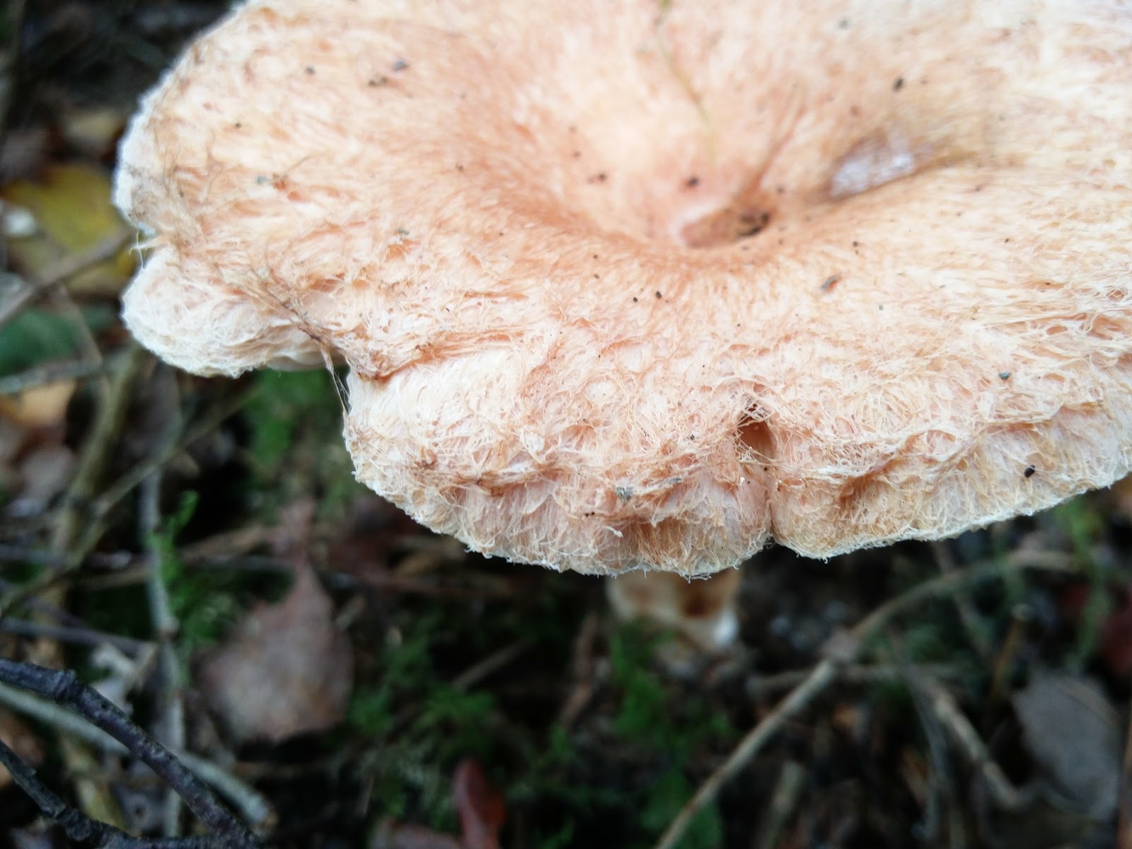
Found these in Frith Wood today, West Sussex. I have no idea what they are.

Sunday 5 November 2017

X-rated

We found this beauty growing on the edge of a woodland near Steyning this afternoon.


We'd seen quite a few Parasol Macrolepiota procera still putting on a show and at first we thought this was another one of those. But the white, slender stem gave it away as something different...


 ... I believe it's the Slender Parasol Macrolepiota mastoidea which I've seen only once before, on a field course in Suffolk with Geoffrey Kibby.

It gets its scientific name from its resemblance to, yup, you've guessed it...


Boobs.

For the record
Date: 5 November 2017

Location: Near Spithandle Lane, Ashurst, West Sussex
Grid reference:  TQ174140

Record entered into FRDBI 07/09/2018

Mushrooms on the verge (2)


I took a quick stroll around the village yesterday; ostensibly to get some exercise (I'm on a health kick) but these days I can't help but keep one eye out for mushrooms.

The big boxy Agaricus were back in Horton Wood but I ignored those. I spent ages looking at them last time they appeared (as evidenced here) and the main conclusion I came to is Agaricus are more difficult to identify than I thought they were. I think I need to get Geoffrey Kibby's book on 'The Genus Agaricus in Britain' to have a proper go at these, but it hasn't quite made it to the top of my fungus-related purchasing wishlist (which keeps getting longer).

But I did stop to look at these, growing on a grass verge. There had been a bit of rain and these were very slippery to the touch. The caps were slightly scaly, especially towards the centre, and around the edges hung obvious veil remnants.


With a few seemingly distinctive features, I thought these might be easy to identify so I took a specimen home for further examination.

It produced a brown spore print.


Since I brought them home yesterday the gills have turned a clay-brown colour and the stem has taken on a rusty colour towards the base.


Entering these features into MycoKey took me towards the Pholiota.

The macro features look quite promising for Sticky Scalycap Pholiota gummosa, as described in Roger Phillips 'Mushrooms'.

The spores look the right sort of shape and size too.

1000x magnification. Spores are around 7 microns in length.

But according to Funga Nordica, Pholiota gummosa should have some quite interestingly-shaped cystidia. I haven't had any luck finding these. All I've been able to find is loads of basidia.


1000x magnification. Stained in congo red.

*has one last look*

Oh wait! What are these...


I think those might be the cheilocystidia I was looking for.

And I this must be one of the "scattered" chrysocystidia.


By jove I think I might be able to confirm this as Pholiota gummosa after all. Go me! (This has only taken three hours.)

I think the next one's easier...


Beige caps again but with a pronounced tan-coloured nubbin (or 'umbo') in the middle.

The gills are free and distant, and produced a white spore print.


I think I'm on safe ground identifying this one as the Fairy Ring Champignon Marasmius oreades.

So, there we go. Turns out it's not just the little brown jobs that can be tricky: it's the medium-sized beige jobs too.

For the record
Date: 5 November 2017
Location: Small Dole (verge adjacent to the Shoreham Road)
Grid reference: TQ214131

Records entered into FRDBI 07/09/2018

Saturday 4 November 2017

A fungus foray at Swan Barn Farm

Sunshine on Swan Barn Farm

I joined Sara Shepley and the West Weald Fungus Recording Group (WWFRG) on Thursday for a foray in the woods around Swan Barn Farm on the edge of Haslemere. This site is managed by the National Trust and the ranger there, Matt Bramich, had kindly offered to take us round. There wasn't much about in the way of grassland fungi, so we headed into the woods.

We found various different brackets and crusts: Birch Mazegill Lenzites betulinus and the much more maze-like Oak Mazegill Daedalea quercina; as well as some very smart-looking Turkeytail Trametes versicolor. We also found a patch of Sulphur Tuft Hypholoma fasciculare which someone said glows quite impressively under UV light – I must try that out some time!

This little group was found growing on well-rotted wood in an area of wet deciduous woodland. They didn't ring a bell with anyone, so I offered to take them home for a closer look.


With no clue as to even what genus these Little Brown Jobs (LBJs) might belong to, I started off with MycoKey which has a pictorial, multi-access key to genus.

The caps looked like they might be minutely hairy.


The gills looked to me to be either shortly decurrent or perhaps slightly emarginate, and they produced a brown spore print.


Entering all these features into MycoKey seemed to take me towards either the genus Flammulaster or Tubaria.

The next step was to look at the spores. Here they are mounted in water.


One sub-unit on that scale is about 1 micron, so these spores are pretty big: up to about 15 microns in length.

Here they are mounted in Melzer's reagent. They've gone a bit brown, but I'm not sure if they've gone reddish-brown enough to be described as 'dextrinoid'.


I think these sort-of almond-shaped spores would be described as 'amygdaloid' which looks completely wrong for Tubaria, so I think we can rule that genus out. It's not looking promising for Flammulaster either as the spores in that genus are generally less than 10 microns long.

On first appearances these mushrooms might look like LBJs but microscopically they've got loads going on! The gill edge is covered in these long-pronged cells with swollen bases: 'urticoid cheilocystidia'. And at the top right here you can see a two-pronged basidium (basidia are the cells which hold the spores).


The cap cuticle ('pileipellis') too has an interesting structure, with chains of elongate cells.

400x magnification.
Here's one of those cells at higher magnification. I think that little bump at the bottom left-hand end might be a clamp connection, linking one cell to the next.

1000 magnification.
I got stuck at this point, so asked for some help on the British Mycological Society Facebook page, here. Richard Shotbolt and Neil Mahler have replied to say the microscopic features look typical of the genus Naucoria which has fairly large, 'amygdaloid' spores; but the macroscopic features don't look typical of Naucoria.

I can't get my observations to fit with any of the Naucoria descriptions in Funga Nordica, so I think I'm stuck again. Might have to give up on this one. Still, nice to have seen these interesting microscopic features.

UPDATE 12/11/2017 Well I decided not to give up on that Little Brown Job after receiving a third opinion from Ken Burgess over on the BMS Facebook page who said my collection matches very well with specimens he has found this season. Ken explained that the spore sizes can be quite variable, and one ought to measure about 20 spores accurately to get a range and an average. He also mentioned that spores from 2-spored basidia are generally larger than those from 4-spored basidia (like piglets from a smaller litter, I suppose), so that could account for the larger spores in my collection.

I got a new bit of kit this week a USB eyepiece camera for my microscope – so I spent some of yesterday afternoon calibrating it so I could get some more accurate measurements of those spores.


I found the spore length to range from 11.1 to 14.2 microns with the average length being 12.8 microns and the average width is 6.1 microns. These average dimensions are *almost* within the range described in Funga Nordica (9-12 microns x 5-6.5 microns).

I've also had confirmation from Sara Shepley that there were Alders around in the part of the woodland where this collection was made, and Nick Aplin has been in touch to say he can see fragments of Alder rootlets in my photograph above. 

I think this gives me enough information to confirm the collection as being Naucoria escharoides, noting the presence of 2-spored basidia and the larger spores.

These Common Earthball Scleroderma citrinum looked like they'd been parasitised by another fungus. Not sure what.


And then I came across some tiny, dusty white fruit bodies growing out of a pupa, nestled among moss on a fallen tree trunk.

Image taken under the stereomicroscope.


I had some help from Nick Aplin of the Sussex Fungus Group on this one and looks like it's Isaria farinosa (formerly Paecilomyces farinosus which is what Barry from the WWFRG said he thought it might be, but I spelt it wrong when he told me so I didn't manage to find a description straight away). It is an entomopathogenic fungus which means it feeds on insects. This individual looks like it's feeding on a moth pupa. Sorry moth fans!

Mike Waterman found a few fruit bodies of this rather lovely-looking Ganoderma.


The pores underneath looked quite big.


And the pore tubes and flesh turned quite yellow when cut.


It was thought this might be Ganoderma resinaceum (TBC).


This neat little round fruit body was growing on a well-rotted fallen branch.


Underneath it had quite large cream pores.


And here's the side view.


The base of the stem looked like it was turning black and it was suggested in the field that this was the Blackfoot Polypore Polyporus leptocephalus. Looking at the photos now, I see that the cap is fringed with hairs...


.. This makes me wonder if it could be the similar species Fringed Polypore Polyporus ciliatus. However, the pores don't look small enough to be P. ciliatus which Geoffrey Kibby, in his book 'Mushrooms & Toadstools of Britain & Europe Vol. 1', says has 5-6 per mm. This collection only has 2 or 3 pores per mm, which is a better fit for P. leptocephalus. Hmm.

The Collins (illustrated) Fungi Guide says that the Funeral Bell Galerina marginata can be "solitary" or found growing in "small tufted groups". But we found it growing great profusion on this rotting tree stump.


UPDATE 12/11/17 ***ERROR ALERT!*** I got that one wrong. Nick Aplin has kindly alerted me to the fact these are Sheathed Woodtuft Kuehneromyces mutabilis. This species often occurs in large tufts like this, on stumps of broadleaf trees, and the stem being scaly below the ring and smooth above is a particular feature of this species.You can see it better in this other photo I took.


This Tricholoma was a new species for me.


It was found growing among Beech trees (my photo isn't from the spot where it was found), which means it looks good for Burnt Knight Tricholoma ustale. Tricholoma are mycorrhizal and there is a similar-looking species which is associated with birch (Birch Knight Tricholoma fulvum).

And finally, this Amanita was nice to find in its prime (almost, except for the chunks the slugs have taken out of it). I thought at first it might be a Blusher Amanita rubescens but it had not a hint of red about it.


The brown cap surface was covered in grey-ish veil remnants.


It was concluded that this is Grey Spotted Amanita Amanita excelsa var. spissa (also referred to as Amanita spissa).

The Collins (illustrated) Fungi Guide notes that Amanita excelsa var. spissa is similar to Amanita excelsa var. excelsa but has a characteristic radish-like smell (I don't remember sniffing it, unfortunately). Apparently, "it is widely believed that var. spissa is the more common variant but var. excelsa is itself a very variable toadstool and the two forms have been greatly confused so records are uncertain". Another corner of mycology which is painted in shades of grey!

For the record
Date: Thursday 2 November 2017
Location: Swan Barn Farm, Haslemere, Surrey
Grid ref: SU9132 and environs

All records to be submitted by Sara Shepley via the West Weald Fungus Recording Group

Friday 3 November 2017

In a copse near Plaistow

I joined Sussex Fungus Group last Sunday for a foray in a copse near Plaistow, West Sussex: a return to this site which I visited for the first time last year (here).

We began with a poke around in the wood piles where we found various crusts and brackets, the usual suspects: Turkeytail Trametes versicolor, Birch Mazegill Lenzites betulina, Purplepore Bracket Trichaptum abietinum, Birch Woodwart Annulohypoxylon multiforme and Bitter Oysterling Panellus stipticus.

Conifer Blueing Bracket Postia caesia was a new one on me, and rather charming.


We also came across the Funeral Bell Galerina marginata growing here and there on bits of rotting wood.


There are lots of Galerina species but the ring around the stem makes this one quite distinctive. We also found this younger specimen, with its particularly neat-looking ring.


Moving on into an area of dense mixed woodland, we found quite a colony of Fenugreek Stalkball Phleogena faginea. I had to get my face right up against the bark to get a whiff of their curry-like scent.


There were masses of these around: Trooping Funnel Clitocybe geotropa.


Here's a rather slug-eaten pair, showing the deeply decurrent gills (or what's left of them).


Growing in an area with Birch, we came across this rather statuesque milkcap which Nick Aplin identified as Wooly Milkcap Lactarius torminosus.


The wooly hairs on the cap and margin are characteristic of this species.


I think we decided this was Brown Birch Bolete Leccinum scabrum.


There was no colour change in the flesh, upon being cut.


In this area of woodland we also came across a lone Slimy Waxcap Hygrocybe irrigata. So slimy I couldn't put it down! (I'll never get tired of this trick.)


I got super excited when we found these tiny orange blobs on a log, because I LOVE these. They're fruit bodies of the slime mould Hemitrichia calyculata.


Luckily Mark Colvin was with us, of Secret Nature fame, and he got this super shot of them. Aren't they gorgeous!

Image © Mark Colvin.

Clare Peters found this Wood Blewit Lepista nuda.


This species has really beautiful lilac colour to the gills when young.


The lilac cap will gradually turn buff-brown with age.


The Collins (illustrated) Fungi Guide describes Wood Blewit Lepista nuda as widespread and very common, but I don't think I've come across it before.

There were lots of Clouded Funnel Clitocybe nebularis dotted around. This species is starting to feel quite familar to me now, as I've seen it all over the place this year.


And an exciting find because I like grisettes was Snakeskin Grisette Amanita ceciliae. The pale grey-brown, white-flecked stem is characteristic of this species.


The cap is covered in dingy grey, warty patches of volva and it has the striate margin which is a feature of the grisettes.


We did manage to re-find the rare Chlorencoelia versiformis which we discovered on our foray here last year (photo here). But it was looking a bit past it.

We also found an amazing little fungus which got dubbed 'Tiny Tube Thing', until Nick Aplin remembered it's called Henningsomyces candidus. There's an article about this species on the Cornell Mushroom Blog (here) which describes the fruit body as looking like miniature macaroni. So true!

For the record
Date:  29 October 2017
Location: Private site near Northchapel

All records to be submitted by Nick Aplin, Sussex Fungus Group