Saturday 25 April 2020

Violet rust is orange


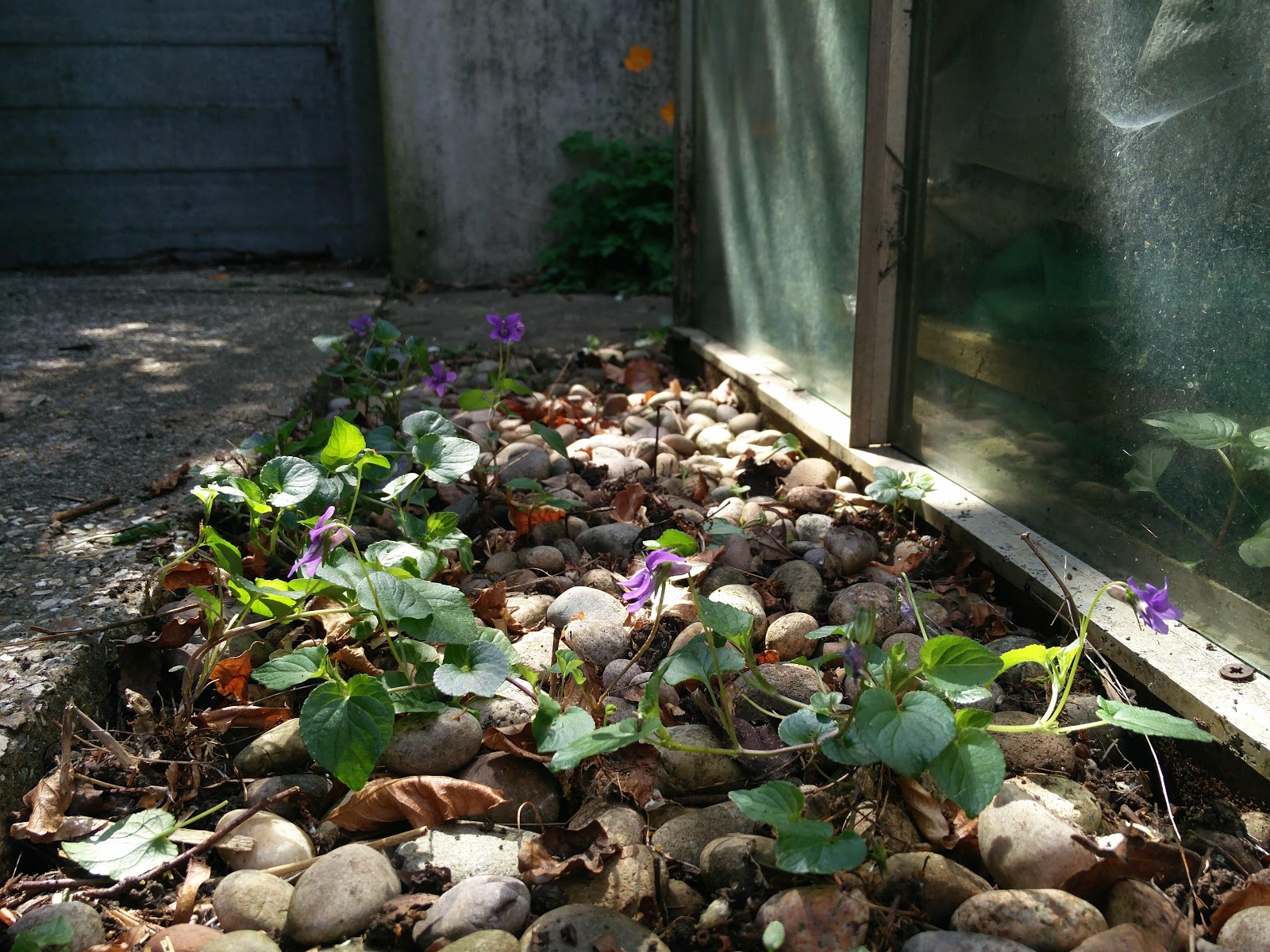
There's a place near my back door where the violets grow. They've seeded themselves there and I am happy to encourage them in this shady strip where nothing much else grows.

Recently I've been reading an old book on 'Rust, Smut, Mildew & Mould' by Mordecai Cubitt Cooke (5th ed., 1886 — you can find it in the Biodiversity Heritage Library, here) and was particularly struck by a description of one species of rust ...
"... which attacks the violet. The sweetest of flowers as well as the earliest, in despite both of its odour and its humility, becomes a victim to one or more of the ubiquitous race of fungi. Thickened spots at first appear on the leaves; the petioles, or the flower stem, or even the calyx, become swollen and distorted; and at length the cluster-cup breaks through."
So I've been keeping an eye on the violets, wondering if I'd catch sight of these so-called 'cluster-cups'. This morning, I found what I was looking for.


Glancing down from above, it was hardly noticeable. But looking closely at each plant, I found the rust was present on lots of them: on the leaves, the petioles and even the calyx — just as Cooke had described.


Correctly identifying the host plant is a critical step in identifying rusts and often the trickiest part for me. I'm no botanist, but when I was a girl my mum used to 'keep things interesting' on a long walk with a promise of two pence for every plant I could name (twenty pence in winter) — so I can get as far as... "dog violet?"

Happily, Sussex Botanical Recording Society has shared a brief guide to Sussex violets, here. And I think the pale upturned 'spur', notched at the end, suggests Common Dog Violet Viola riviniana.


Scientific knowledge and taxonomy of rusts has moved on somewhat since 1886, so — armed with a hopefully-accurate ID on the plant — I consulted a more modern tome to identify the rust. I've just recently acquired a .pdf of 'Dutch Rust Fungi' (from here) and it includes helpful keys as well as descriptions and illustrations, in English as well as Dutch. I conclude that this 'cluster-cup' — the 'aecial' stage in the rust's life cycle, also known as stage I — is Puccinia violae.

Here is a close-up of those beautiful little 'cluster-cups' on the underside of a leaf. 


The book on 'British Rust Fungi' by Wilson & Henderson (1996) includes a more detailed description of the 'cluster-cups', or aecia [singular = aecium], of P. violae. The outer wall of the 'cup' which you can see here is known as the peridium and is described as having a "white torn revolute [meaning 'curled back'] margin", which you can see here in a focus-stacked image of my collection.


The orange mass inside, and the tiny orange specks scattered about on the leaf's surface, are the aeciospores. Poised to infect their next victim... — if you frame it in Cooke's terms. Or just being aeciospores... — if you're inclined to be less judgemental about this thing called life

I don't think there is a 'Spotters Guide to Rusts' but, if there was, this species would surely be in it as it's apparently a common one to find. Ingram & Robertson mention it in their book on 'Plant Disease' (1999) and it's an example of a rust fungus which lives out its whole rather elaborate life cycle in association with one species of plant, in this case Viola riviniana, so it's known as 'autoecious'. There are photos of the rust's other life stages on the Plant Parasites of Europe website, here. So naturally I'll be looking out for those! Although they're rather less showy.


(I've got another rust in the garden which spends part of its life on my Pendulous Sedge Carex pendula and part of its life on something else — possibly the nettle patch or my fruit bushes — but I'm still trying to get to the bottom of what's going on there, so will have to save my 'heteroecious' rust fungus for a future blog...)

For the record
Date: 25/04/2020
Location: My garden, West Sussex

Saturday 18 April 2020

Lockdown foray

I'm taking part in a 'lockdown foray' today with other folks over on the British Mycological Society Facebook page, in memory of Richard Shotbolt.

Here's this morning's finds from my garden in West Sussex.


An aging patch of garden bluebells provided my first find...


... the distinctive, elliptically arranged pustules of Bluebell Rust Uromyces muscari.


Tucked away at the back of the garden, a self-seeded tutsan (Hypericum sp.) was showing telltale yellow spots on a few of its lower leaves.


Tutsan Rust Melampsora hypericorum seems a likely identity for this one. I recall I found something like this before at Rowland Wood, here.


I think I've got something similar on the Petty Spurge Euphorbia peplus that grows in the cracks on the patio: Melampsora euphorbiae. I've recorded this in the garden before, here.


Then the plant identification gets a bit challenging... The tiny white flowers on this plant have six stamens and I think it might be Wavy Bittercress Cardamine flexuosa.


Whatever it is, it's got some impressive white blisters on its leaves.


Looking in 'Microfungi on Land Plants' by Ellis & Ellis, I'm thinking this must be the 'white rust' Albugo candida. But it's not a fungus! It's an oomycete. So I lose a point there.

Next up I've got some fuzzy-looking spots on the underside of a cultivated primrose.


From the top, you can just make out some brown spots and the leaf is yellowing around them.


I'll be honest, I don't really have any idea what this is. Is this what downy mildews look like? Kinda fuzzy-looking?

I can see loads of what-I-assume-are fungal hyphae. But I don't think I'm anywhere close to an identification with this one.


I had a couple of things on some dead currant twigs.

There was this black, bristly looking thing. I thought it might be Chaetosphaerella phaeostroma, which I've come across before (description here). But all I'm seeing is black bristly stuff, and no fruit bodies which might help me confirm an identification. Hmm.


These tiny yellow specks looked like they must be something fungal. 


They are miniscule cup funguses!


Not sure how to progress towards an identification with these, as I think there are a few different genera that can look like of like this: e.g. Bisporella, Calycina and Orbilia?

Finally, I've got this poroid resupinate fungus which was growing on the underside of a plank that was lying on the ground.


Hmm. I've been flicking through 'Fungi of Temperate Europe' and there are rather a lot of species that look a bit like this. Might give up on this one!

For the record
Date: 18 April 2020
Location: My garden, West Sussex

Friday 17 April 2020

Rust Quest

'Rust Quest' was a bit of nonsense I came up with last summer, to add a little excitement to my fungus forays. This was one of my more memorable finds – on the patio! – just shared on Twitter at the time.


Now seems like a good time to seek out a few more rusts and smuts around here!

For the record
Date: 9 July 2019
Location: Small Dole, West Sussex

Monday 13 April 2020

Crystals in the leaf litter

Another day, another poke about in the garden.

This dead bramble leaf provided much distraction today. More specifically, it was the tiny grey blobs on the bramble leaf that caught my eye. See 'em?


Once I'd got them under the stereomicroscope, I could see that they were the 'fruit bodies' [sporangia] of a slime mould. So I got my slime mould book out: 'The Myxomycetes of Britain & Ireland' by Bruce Ing. I don't have much experience with slime moulds, but I guessed this one might be a Physarum species...


The sporangia were just stunning to look at under the microscope so I spent a while getting a focus-stacked image of one. I saw that the outer surface [peridium] is covered in frost-like particles.

Focus-stacked image of the peridium. Around 40x magnification.

The round mass of spores, enclosed by the peridium, sits on top of a stout white, slightly furrowed stalk – shown here on one of the sporangia which had neatly broken into cross-section.

Focus-stacked image of a sporangium in cross-section. Around 40x magnification.

You can see here the round flat base to the stalk which anchors the sporangium to the surface it's growing on: the hypothallus. From the tip of the stalk [the columella] tiny threads radiate outwards; these threads form the capillitium: the structure which holds, and assists dispersal of, the spores.

Here are a few of those threads under the microscope.

Section of capillitium. Mounted in water, 400x magnification.

Flicking through Ing's book, the description of Physarum leucopus sounded close to what I was looking at. But it noted also a "close resemblance to Didymium squamulosum".

I dipped into the British Mycological Society Facebook page, to see if previous discussions there would illuminate the difference between these two species... And I found that Steven Murray had commented on a similar-looking collection last year (here) that angular star-shaped crystals are indicative of Didymium.

I scanned across the material I'd prepared under the microscope. And I think I've found what I was looking for.

Star-shaped [stellate] crystal shown with two minutely-warted spores. Mounted in water, 1000x magnification. Focus-stacked image.

Wow! Slime moulds really blow my mind. How do they create crystals out of bits of old bramble? Damned if I know.

I think that makes my collection Didymium squamulosum.

For the record
Date: 13 April 2020
Location: My garden, West Sussex

Saturday 11 April 2020

The Joy of Sticks

One unexpected consequence of our current circumstances is that my garden – the location for Michael Blencowe's daily 'Corona Wildlife Diary' – has become ever so slightly famous.

It's been a joy to see people sharing stories of life, wild life, continuing on around us. But when the camera turned to my back garden, I confess to a sudden and powerful empathy with Hyacinth Bucket and that matter of 'Keeping Up Appearances'. Because – with time taken up by work and other interests – our garden had become a bit of a mess.

So it was that I found myself on the first day of the bank holiday intent on tidying up the 'vegetable patch'.


Last year's Fennel has stood here all winter; its stems now well and truly dead. So I thought I'd start by clearing these away. It took longer than I thought as, while I was chopping them into bits to go in the garden waste bin, I got rather distracted by the tiny bumps and speckles which adorned them.

The thin stems in the Fennel's upper reaches were most intriguing, being covered in a smattering of tiny fuzzy-looking specks. So some of these were saved from the garden waste bin and came into the house with me. I popped them in a box with some damp kitchen roll and waited to see what would happen.


Here's one of those stems after it's moistened up a bit. You might just be able to make out those fascinating specks?


Another hour or two passed, while I figured out how to change a light bulb in the stereomicroscope.

It was worth it, because these aren't just any old specks. They are in fact beautiful soft grey cup funguses fringed with crystal-white hairs.


At the highest level of magnification, I could just make out some tiny square clusters of dots – a telltale sign that what I'm looking at here is a fungus which holds its spores out on prongs [sterigmata]: it's a basidiomycete.


I think what I've got here might be Lachnella alboviolascens; but there may be similar species and I haven't yet managed to track down a key.

The authors of 'Fungi of Temperate Europe' describe Lachnella as "a genus of dessication-tolerant cyphelloids... In dry weather the fruit bodies close and form small, hairy balls." You can actually see this in action under the microscope – these photos were taken over the course of about 15 minutes.


Those weren't the only interesting specks on the Fennel.


The thick stems were covered in tiny black bumps which – taking a lead from 'Ellis & Ellis' – I think might be Leptosphaeria libanotis, with a "depressed-globose" shape.


I can't work out if the even-tinier specks shown here [TOP] with the Lachnella for scale [BOTTOM] is a baby version of the Lachnella, or something different. Might need to get the slide microscope out...


Anyway, I think we've established why the garden's such a mess. And maybe messy's just fine for funguses.

For the record
Date: 11 April 2020
Location: My garden


From https://www.thepunctuationguide.com/en-dash.html
© 2020 thepunctuationguide.com


From https://www.thepunctuationguide.com/en-dash.html
© 2020 thepunctuationguide.com


From https://www.thepunctuationguide.com/en-dash.html
© 2020 thepunctuationguide.com


From https://www.thepunctuationguide.com/en-dash.html
© 2020 thepunctuationguide.cas the location for