I joined
Sussex Fungus Group on a foray at another Sussex Wildlife Trust reserve this morning:
Ditchling Beacon. We were joined by Sussex Wildlife Trust ecologist
Graeme Lyons, who took us on a tour through the site's different habitats
— including several patches of species rich chalk grassland.
An area Graeme called 'the horseshoe', around TQ326134, provided quite a bit of interest. Here and there the sward was dotted with small inkcaps, all in various stages of deliquescence.
They were so small, I didn't fancy my chances at getting one home and keying it out. Although I do keep meaning to have a go at using
Derek Schafer's key to 'Ink Cap' type things...
Nearby, a cluster of dark brown
Panaeolus, with suggestions of a hygrophanous cap.
Nick Aplin pointed out how the darker brown gills with white, sterile edges helps with differentiating
mottlegill Panaeolus from similar-looking
conecap Conocybe species. I think this one's
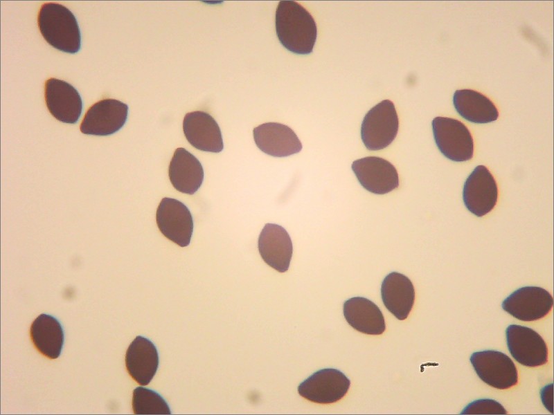
Turf Mottlegill Panaeolus fimicola.
The spores are smooth and around 11.5 - 13.5 microns long and 7.5 - 9 microns wide {based on 10 measured}.
 |
| Spores at 400x magnification in water. |
But I had a devil of a time trying to locate the sulphidia which are supposed to be present on the gill face. Couldn't see any when I did this gill trama thin-section...
 |
| Gill trama thin-section at 100x magnification in water. |
So I resorted to doing some squashes of the gill face. I've half convinced myself that I've found a sulphidium in this image. But I'd be happier if it was more yellow, and less brown.
 |
| Gill face squash at 400x magnification in water. |
The cheilocystidia, on the edge of the gill, look about right for
P. fimicola.
 |
| Gill edge squash at 400x magnification in water, showing cheilocystidia. |
After the
Panaeolus, we found an absolutely perfect
Wood Blewit Lepista nuda. Those dusky-mauve gills are just gorgeous.
Next to those, a rather more characterful
Panaeolus:
Petticoat Mottlegill Panaeolus papilionaceus. If you catch these when you can still see that ragged veil, like bunting around the bottom, then I think I'm right in saying they're identifiable in the field.
Around the corner we picked up our first waxcap of the day:
Toasted Waxcap Hygrocybe colemanniana.
This species was a new one for me. The distinctive cap colour and
decurrent, intervenose gills, make it identifiable in the field. We found some more fresh specimens in the old quarry at the bottom.
The old quarry proved pretty productive for waxcaps and we lingered here a good while.
 |
| Looking for waxcaps in the old quarry. |
These were identified as
Snowy Waxcap Hygrocybe virginea, as they had no particular scent.
Reading through
Boertmann's 'The genus Hygrocybe', I note that their field characteristics would fit his description for
Hygrocybe virginea var.
fuscescens — which he says is "similar to H. virginea var. virginea except for a brownish central spot on the cap and a more or less distinct, brownish, radial striation." He also notes that it is "apparently always on calcareous or basic soils in grasslands, fixed dunes and road verges." Which would fit with our collection.
These
Blackening Waxcaps Hygrocybe conica looked like they had showed up late for a Halloween party, with those stunning orange colours.
Now I've had a chance to re-read Boertmann's description, I'm wondering if this stockier, yellow one would fit with
H. conica var.
chloroides.
Graeme also found a nice patch of yellow (not blackening) waxcaps, which I think keyed out as
Golden Waxcap Hygrocybe chlorophana.
Poking around in the rough vegetation, where the grassland meets the surrounding woodland, we found a couple of interesting things, including this
Redleg Club Typhula erythropus on a fallen Ash petiole (?).
From there we headed back up the hill through the woodland and rougher grassland on the eastern side of Ditchling Bostal.
We had another
Typhula-type-thing growing on dead ivy stems, in a log pile. Nick Aplin took this one away to ID, along with another one we'd found growing on dead
Helianthemum stems.
I volunteered to attempt an ID on this
Crepidotus, growing on a fallen branch of Ash.
It has broadly ellipsoid to globose spores, around 7 microns long x 6 microns wide (average Q value = 1.2) which are covered in small warts.
 |
| Spores at 1000x magnification, in water. |
And I think I can just about make out some simple, unbranched cheilocystidia here.
Using Funga Nordica (2008), this keys out to
Flat Oysterling Crepidotus applanatus.
This
Pluteus was growing on an old tree stump.
The striate margin to the cap and wrinkled centre struck me as different to your usual brown
Pluteus.
It has broadly ellipsoid to globose spores, averaging 7.3 microns long x 6 microns wide (Q value = 1.2).
This is what the cap cuticle looks like. A cellular structure?
 |
| Cap cuticle near centre, 100x magnification in water. |
 |
| Cap cuticle near centre, 400x magnification in water. |
I've spent hours trying to satisfy myself I've located the pleurocystidia. I found this pale shadow of a thing, in a preparation stained with PlaqSearch.
 |
| Pleurocystidium? 1000x magnification in water + PlaqSearch. |
And eventually found these similar-looking things in a preparation stained with Congo Red.
 |
| Pleurocystidia? 400x magnification in water + Congo Red. |
If these are indeed pleurocystidia, then
– following the key in Funga Nordica
– would lead me towards
Wrinkled Shield Pluteus phlebophorus. But I'm not sure the cap cuticle is right for that species (I can't find a good photo to compare with online).
I got very confused and disorientated while searching for cystidia! I found billions of basidia, and a few cylindrical to lageniform cells, which I thought could be cystidia. Or perhaps just immature basidia
— how would I know?
 |
| Basidium and possible cystidia. 1000x magnification in water + PlaqSearch |
 |
| Sausage-shaped thing. A cystidium? Who knows. 1000x magnification in water + PlaqSearch |
I did also consider
P. podospileus, but discounted it on account of the stipe not having "numerous
dark brown punctate floccules"
– although we did comment upon the stipe being somewhat floccose, when we collected it. Richard Illiffe's key in Field Mycology indicates there are several other brown-capped
Pluteus species which have a preference for calcareous woodlands and have this feature of being translucently striate when moist, with a cellular cap cuticle, including:
P. cinereofuscus and
P. satur (=
pallescens). But I haven't managed to satisfy myself they are a match for what I've found.
So I think I'm stuck with that one
— guidance appreciated!
I also found myself drawn in by several
Ascocoryne specimens, over the course of the foray.
Here we found an
Ascocoryne anamorph and teleomorph growing together. The distinctive anamorph makes this one
A. sarcoides.
I also picked up the teleomorph of another
Ascocoryne, with a thinner, more cartilaginous 'cup'.
I see there is a
'Key to Ascocoryne' available from the asco-france website, by H. O. Baral. This explains that
Ascocoryne species in section 'Sarcoides' have 2 (-4) large oil droplets within the mature ascospores.
I thought I'd check this on our collection of
A. sarcoides. Yup!
 |
| Mature ascospores from A. sarcoides. 400x magnification in water. |
The spores from my other collection are completely different, with many small oil droplets. They look exactly right for section 'Cylichium'.
Here's a closer look at those ascospores.
I think I can probably get away with calling this collection
A. cylichnium.
Finally I should give a mention to the properly stinking patch of
Stinkhorn Phallus impudicus which we passed on our way back up the hill.
The flies were enjoying this one.
For the record
Date: 10/11/2018
Location: Ditchling Beacon
All records to be submitted via Sussex Fungus Group