Friday 8 May 2020

Kuehneola uredinis (or not?)


This orange-streaked bramble stem caught my eye today. 

Bramble Rubus fruticosus agg. hosts a few species of rust fungi but, as far as I know, only one which looks like this. These longditudinal pustules are the uredinia (stage II) of Kuehneola uredinis.


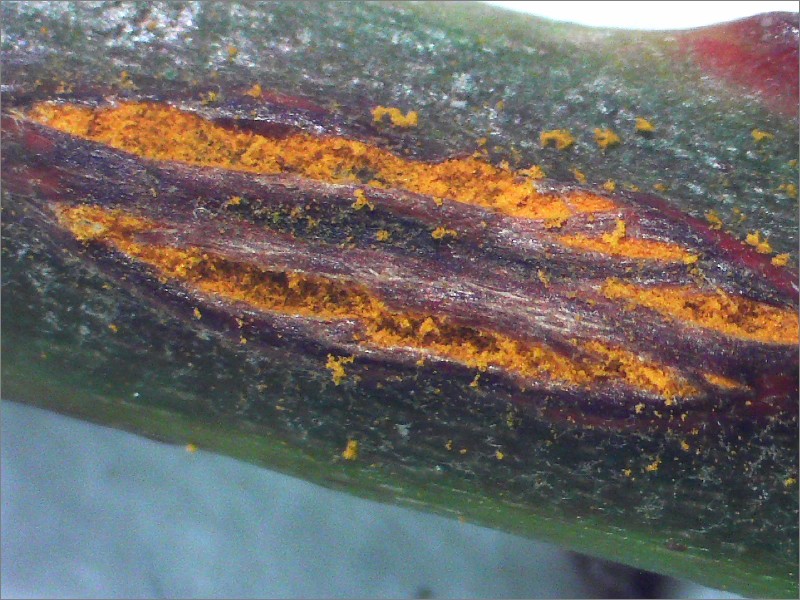
The UK species inventory lists its preferred common name as Pale Bramble Rust, with Bramble Stem Rust also in use. But they're both rather unsatisfactory. At this stage in this species' life cycle you can see it's very much not 'pale'. And at other stages of its life cycle you'll find it on growing on the bramble leaves, not the stem. So I'm inclined to forget the common name for this one.

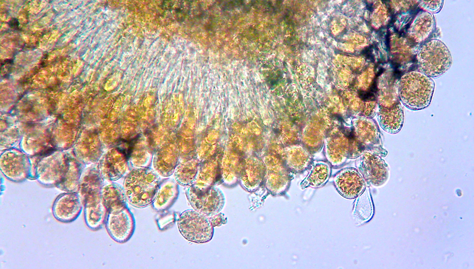
I think you can get away with identifying this species from its field characters, but I did have a look at the uredospores.


I think this is what they're supposed to look like! I guess that's an immature one on the left, with a stalk still attached?

UPDATE 10/05/2020

OK - turns out this one isn't quite as easy as I thought. Apparently Phragmidium bulbosum can also look similar in the field and occur on bramble stems.

Differences between P. bulbosum and K. uredinis sound rather subtle to me (with reference to Dutch Rust Fungi). 
  • P. bulbosum should have paraphyses, but I can't figure out what they'd look like. 
Is that stalked thing in my image above a paraphysis? I'm inclined to think it's a spore; I saw reference in Wilson & Henderson's 'British Rust Fungi' to Phragmidium spores being "borne single on pedicels" - so perhaps the stalk-looking thing is a 'pedicel'?
  • P. bulbosum uredospores are 'distantly echinulate' whereas in K. uredinis they're 'densely and finely verrucose/echinulate.
This is the best illustration I can find of that distinction:

Reproduced from 'British Rust Fungi' by Wilson & Henderson (1966).
Incidentally, Grove (1913) seemed to have a completely different view on what K. uredinis spores look like.


Wilson & Henderson treat Grove's K. albida as synonymous with K. uredinis; but that uredospore illustration in Grove looks suspiciously like Wilson & Henderson's illustration for P. bulbosum. Hmm... Confusing!

The Plant Parasites of Europe website has images of K. uredinis uredospores (here) but no comparable photos for P. bulbosum.

I got this other micrograph of my collection, which shows something spotty, i.e. a 'verrucose' or 'echinulate' uredospore? I don't know why it's clear (hyaline) – did the fungus produce an empty spore for some reason? Maybe the other ones look like this too but you can't see the details of the uredospore wall usually, against the orange contents...?  

  • Also P. bulbosum is described as 'yellow' and K. uredinis is 'orange-yellow'. Well, I would describe my collection as 'orange-yellow' but that's not much to go on.

Anyway. I've read everything I can think of to read, and googled everything I can think of to google and I'm still not sure what I've got here. Might have to ask an expert.  

UPDATE 25/05/2020

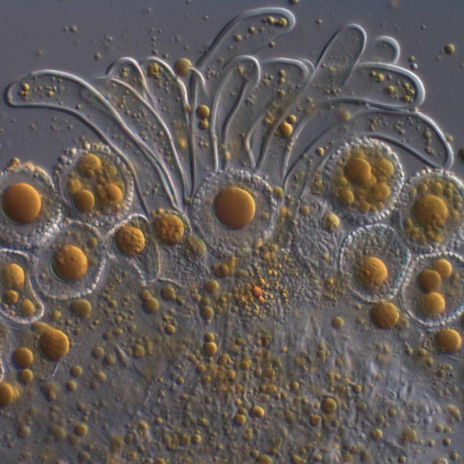
OK - so I feel like I left this one kind of unfinished... And the help I received in the comments here and via the British Mycological Society Facebook page have spurred me on to have one more look at this bramble stem rust.

Brian Douglas let me know that the paraphyses of Phragmidium should look something like this. And I was advised to look at a cross-section through the stem rust pustule, to have a proper look for them. So that's what I did. Here is a shapshot from one of the cross-sections I looked at.


I'm now pretty sure my collection doesn't have paraphyses. So I'm going with an identification of Kuehneola uredinis.

I've learned a few other things along the way. The clear structure I was talking about earlier is a dead spore. You can see another one in the image above.

The growing spores are held on a stalk called a 'pedicel' - and that is what I was looking at in my first micrograph.
 

For the record
Identification: Kuehneola uredinis
Date: 8/5/20
Location: Home, West Sussex

Thursday 7 May 2020

Withered grass


I think I mentioned the other day I've been reading 'Rust, Smut, Mildew & Mould' by M.C. Cooke (5th edition,1886). It's a terrifically engaging introduction to these microscopic fungi.

Whereas modern texts on 'plant disease' can be rather dry and scientific in style, 'Rust, Smut, Mildew & Mould' presents the study of these fungi as a thrilling voyage of discovery.
"We might traverse the primeval forests of the new world, and explore the unknown regions of the old, and not encounter so much to excite our admiration, or cause our wonder, as lies about our feet at home; marvels which we tread beneath our feet, or kick from our path, because they appear to be only rotten sticks, withered grass and decaying leaves. All this may appear as the dream of an enthusiast, or the ravings of one on whom the moon has shone too often. When Columbus spoke of a new world beyond the seas, which longed to seek and explore, some believed him duped, and others called him mad. We write of no chimerical El Dorado, we speak of no undiscovered world, and yet we seem to allude to wonders still unknown, because so few have had the courage to venture upon the journey for themselves."
Cooke's book ran to six editions and must have encouraged many readers to pursue detailed studies of these microscopic fungi – discovering important economic applications for this knowledge along the way. Nowadays, plant pathology is primarily the preserve of academic and agri-industry types. For the amateur it seems a relatively obscure branch of mycology, in the UK at least. Unless you happen to live in Wales, where there is a thriving 'rust study group' – responsible for producing a number of attractive publications which provide a modern stepping-in point to the study of rust, smut and mildew:
  • Rust Fungus Red Data List and Census Catalogue for Wales (here)
  • Smut and Allied Fungi of Wales (here)
  • Downy Mildews and White-blister Rusts of Wales (info here)
One (not-insurmountable) barrier to studying these fungi in any detail is the need for a microscope and knowledge of how to use it. In Cooke's day, amateur microscopy was quite the craze. He himself was a founding member of the Quekett Microscopical Club (which still exists today, I wanna join!) and would spend evenings at the microscope with fellow enthusiasts.

Microscopists (left, Cooke; right, Berkeley) examining the potato blight fungus. From a cartoon by W. G. Smith, Graphic, 1875. Reproduced from the biography of Mordecai Cubitt Cooke by Mary P. English.
Any long-time readers of this blog will know that the road to basic competence with a microscope has felt like a long one for me. As a 21st century amateur mycologist, opportunities for practical instruction are relatively few and far between. But when they do come along, it makes such a difference having someone actually show you what to do.

I now find myself in the fortunate position of having a good microscope and some knowledge of how to use it. So when my birthday came around, under 'lockdown', I thought I'd treat myself to a new eyepiece camera and a few other bits...


... and I headed out into the back garden, in search of "withered grass and decaying leaves".

I didn't have to go far. The Sweet Vernal-grass Anthoxanthum odoratum in our 'mini-meadow' was covered in rust.


The rust appeared to be 'epiphyllous' – growing only on the leaves...


... forming these scattered orange-brown spots. I fathomed that these must be the 'uredinia' of a Puccinia species; stage II in the rust fungus's life cycle.


You can see how the uredinia have burst through the leaf's surface. Look closely and they're made up of masses of uredospores.


Here are some of those uredospores at 400x magnification. Mounted in water so you can see their true colours.


I didn't know what these wiggly things were. But reading around a bit, I figured out they're paraphyses. The presence of paraphyses, and their shape, is significant when it comes to identifying these rusts.


There's a good, up-to-date, book on rust fungi, 'Dutch Rust Fungi', which the authors have made available online, here. This gives two species of Puccinia occurring on A. odoratum: P. graminis and P. brachypodii. The presence of thick-walled paraphyses, as seen here, suggests P. brachypodii s.l..


The description in 'Dutch Rust Fungi' states that "taxonomic interpretations of this species are manifold" and other authors recognise different species. Elsewhere, I found a description of P. poae-nemoralis on the Plant Parasites of Europe website (here), which seemed to match my observations well, with reference to "hyaline paraphyses that bear a distinct head and often are sharply bent just below".

I consulted with the experts on the UK Fungi forum and received a very informative response from Paul Cannon (here) – providing some additional insights into the complicated world of rust biology and taxonomy. He advised recording this observation under the name P. anthoxanthina (in the sense of Klenke & Scholler's 'Pflanzenparasitische Kleinpilze', 2015); although in the Checklist of British & Irish Basidiomycota this name is considered to be synonymous with P. brachypodii var. poae-nemoralis (so I was on the right track!) and P. brachypodii var. poae-nemoralis is preferred.

It is easy to get hung up on names and worry about selecting the 'right' one, or – horror! – giving your collection a wrong one. The way I see it, a name is just shorthand for a suite of characters observed. Names are useful, as a way of accessing information on people's understanding of nature. But the observation is the important bit: make a good record of your observations and let this be the truth of what you saw. A name is just for our own convenience: a folder in which to file ones observations.

But that's easy for me to say. I am a very slow mycologist; content to wonder at the marvels which we tread beneath our feet.


For the record
Date: 3/5/2020
Location: Home, West Sussex