 |
| Larch Bolete Suillus grevillei popping up in the grounds at Waterside, Cornwall. |
I joined the British Mycological Society (BMS) a couple of years ago, when I realised my interest in fungi was turning serious. From the outside, it looked slightly daunting. There was a form. And I had to find someone to vouch for me. But I figured it'd be worth it to get access to their Field Mycology magazine and keep up with goings on in the fungus world.
Once I'd joined, I discovered the BMS organise a bunch of interesting events. I went along to the Autumn Meeting at Kew, and I booked myself onto the Fungal Identification Skills Workshop in Barnsley, led by Carol Hobart. I found these really useful for putting my local fungus finds into context and developing the skills to delve deeper into mycology.
This year – armed with my student microscope and some basic knowledge of how to use it – I decided to book myself onto the ultimate BMS event: the Autumn Study Week. A whole week of fungus foraying and identification.
The Autumn Study Week is held in a different place every year and this year it was in Cornwall, based at Waterside near Bodmin. There's always a tutor who leads a discussion after dinner about each day's finds; this year it was Jens Petersen, author of that absolutely gorgeous book: 'The Kingdom of Fungi'.
Late September / early October in Sussex had been so dry, I wasn't sure how much fungi we'd find down in Cornwall. But I needn't have worried. Walking from my chalet to the workroom on the first afternoon, I spotted a patch of Larch Bolete Suillus grevillei and from that moment on I was immersed in mycological marvels from morning 'til night – for over five days straight.
I reckon if I can remember five things from a day's foraying, then I'm doing well. So here's 25 things I learned on the BMS Autumn Study week...
1. Want the world on a stick? Here you go...
Our first foray was was at Breney Common where I found this stick.
It didn't look like too much at first. A lemon yellow crust fungus with a party trick: add a drop of potassium hydroxide (KOH) and it turns bright purple. Stuart Skeates identified it for me as Mycoacia uda.
But get it under the stereomicroscope and a heavenly world is revealed, of wide yellow plains and impossible peaks.
Stunning.
2. Hebelomas do have features
Hebeloma is one of the groups of brown mushrooms I usually ignore and walk past. But it was nice to be introduced to the slightly sticky Bitter Poisonpie Hebeloma sinapizans by Richard Shotbolt, who showed me the 'wick' of cap flesh which hangs down into the stipe cavity.
3. This is that thing that Lactarius tabidus is supposed to do!
After several failed attempts recently at coaxing enough milk out of a milkcap to wet a bit of tissue, to see if it was the Birch Milkcap Lactarius tabidus, it was nice to finally get a positive result on this milkcap I found at Breney Common.
4. Helvella macropus is the cutest
I was very taken with these stalked cup fungi. Following Brian Spooner's Helvella key took me to Felt Saddle Helvella macropus.
5. Inocybes: It's all about the base (and the stipe)
Another attendee on the Fungus Study Week was Penny Cullington, author of 'Keys to British Species of Inocybe' (which I obtained on request via Bucks Fungus Group); so I thought I'd take the opportunity to get some identification tips...
Penny explained that the 'pruinosity' of the stipe is a key feature in this group of fungi. You can sometimes see this with a hand lens, but it can also be checked microscopically by looking for caulocystidia and determining whether these are present along part, or along the full length of, the stipe. This is best done from fresh material, and Penny showed me how to prepare a super-thin strip of the stipe surface, for examination down the microscope.
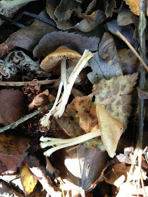
This came in handy after our first foray, as I had found what I guessed was an Inocybe at Breney Common, growing with hawthorn and birch.
Unfortunately, as you can see in the photo, they kind of came apart in my hand the moment I touched them. This meant I missed another key feature I should have been looking for: the nature and shape of the base. Luckily Penny was able to tell me the base of these particular specimens would have been bulbous ('emarginate').
After Penny's tuition, I managed to get a good look at the thick-walled caulocystidia, and spotted a few nodulose spores sitting on the stem surface.
 |
| Caulocystidia with one solitary spore. 400x magnification in water. |
I made rather a hash of working through the key, but with a lot of help from Penny I eventually got to Inocybe mixtilis. (Hope I've got that right...)
6. Mycocam is really neat
I'd bought a new laptop for Fungus Study Week, just so I could use my microscope camera. But then I couldn't get the camera software to work (argh!), so I thought I'd try Richard Shotbolt's MycoCam 4 software instead...
It was really quick to install, detected my camera straight away and happily I'd remembered to bring my stage micrometer, so with a bit of help from Richard I soon had the calibration set up.
MycoCam is designed specifically for mycology – so it makes measuring spores etc really easy, while still allowing you lots of control over camera settings and image processing. I much prefer it to the software that came with my camera. I won't be going back!
7. A good year for piggybacks (Asterophora sp.)
At both Breney Common and Red Moor the woodland floor was dotted with dead, blackened Russulas. On almost every patch we found the fruit bodies of Silky Piggyback Asterophora parasitica and Powdery Piggyback Asterophora lycoperdoides.
In once place I found what looked like the two species growing together, which made me wonder if I was in fact looking at two different stages of A. lycoperdoides: before and after the chlamidospores had formed on the surface.
(I meant to check the gills, but got distracted by other things.)
8. Mycologists can't resist a two-for-one offer
Once Simon Harding had pointed out these small black things poking up through the moss, I suddenly started to see them all around me, on the mossy lakeside banks at Red Moor. They are Snaketongue Truffleclub Tolypocladium (= Cordyceps) ophioglossoides. A new species for me.
Golden threads connect this parasitic fungus to its host: a buried truffle, from the genus Elaphomyces. (I'm sure Carol Hobart got an ID for the truffle, but I didn't catch it.)
Fascinating.
9. How to sort out those big white ones
As well as dead, blackened Russulas, the woodland floor at Red Moor was scattered in places with big white funnel-shaped mushrooms.
These were identified as Fleecy Milkcap Lactarius vellereus. I've tended to get a bit confused about mushrooms that look like this, but I was told if it produces milk then it's either L. vellereus or L. piperatus – and the latter has a very hot peppery taste. And if it doesn't produce milk (and it's massive) then it's probably Leucopaxillus giganteus.
I really should try and get over my aversion to tasting mushrooms...
10. All the hours in the day is not enough hours to study fungi
I returned to base with various collections, and every intention of studying them all in more detail. But there were too many, and too little time. The possible Psathyrella, Naucoria, Tubaria, etc, never got to being identified. I told myself I'd collect less tomorrow...
11. There is treasure in the dunes
In a sheltered hollow, on the northern side of some dunes, we found some waxcaps poking up through the sward.
I haven't looked at waxcaps much before and I was amazed by their vibrant yellow and orange colours, in the stipe as well as the cap.
The young caps were glossy and slightly viscid, with free gills.
Following Boertmann's key in 'The genus Hygrocybe - 2nd revised edition', it looks like these collections are all Persistent Waxcap Hygrocybe acutoconica (= Hygrocybe persistens). Lovely to see!
12. Blackening Waxcaps: complex
A little further inland, on the coastal grassland, I came across some other waxcaps.
The orange caps had bright reddish tones, and the fruit bodies began to turn quickly black upon handling. Blackening Waxcap Hygrocybe conica? But so different to the ones I've seen before.
I note that Boertmann says, "the whole conica complex is in need of molecular studies and a thorough revision". So perhaps the considerable variation in H. conica hides many cryptic, phylogenetically and ecologically distinct species... Time will tell.
13. Fungi are also available in neon
In the afternoon we headed to an area of scrappy woodland on the edge of Portreath: on a twitch! I couldn't really go to Cornwall without seeing Favolascia calocera: the orange ping-pong bat. And Pauline Penna had offered to show us exactly where to find it.
After being initially underwhelmed by some dried-out specimens near the entrance, I was completely wowed by these fruit bodies, found by Peter Smith.
Even though I knew what we were looking for, I wasn't prepared for them being... Quite. So. Orange.
14. Good things can come in non-descript packages
In the same woodland (near the Flaviporus brownii, which Alick Henrici has written about in Field Mycology) I came across this initially rather non-descript, flappy-looking thing, on a well-rotted fallen trunk.
I wasn't sure what to make of it, but Brian Douglas from the Lost & Found Fungi Project immediately clocked it as a Fungus of Interest.
I'll share more on the identity of this collection in due course...
16. The frustrations of measuring tiny spores (and why I want a new microscope)
As a little teaser for all the asco-fans out there, I will just mention that identifying the fungus above involved measuring a lot of very tiny ascospores.
I was happy to establish, on the fungal identification skills workshop last year, that my microscope is fine (it's basically a 20-year old version of Brunel's SP03 student microscope). And I was pretty happy to get this quality of image out of it, at 1000x magnification under oil immersion.
 |
| An ascospore, about 2.5 microns long. |
And when you've got to faff around popping your camera in and out of the eyepiece all the time, trying to find what you're looking for and get things into focus. And you're fiddling with a desk lamp trying to get the light right. And the heat from the bulb keeps drying your slide out every 5 minutes. And you've used up a whole day and a half of your life that you won't get back. It does rather make you want a better microscope.
(I'm thinking trinocular, with LED lighting and space for four objectives – so if anyone's got any recommendations...???)
15. Imagine you're a tiny chef!
Just when I thought I was done with measuring ascospores (I enjoyed it really 😉), I found another interesting little cup fungus. A flesh-coloured thing growing on a well-rotted bit of fallen dead wood.
Brian Douglas had shown me how to prepare thin-sections under the stereo-microscope with a razor blade – like those tiny cooking videos on youtube! (Which I love.) So this gave me some more material to practise on.
Much like tiny cooking, cutting thin-sections requires tiny utensils, and another great tip from Brian was to use a hypodermic needle to move the bits around: super-sharp with a tiny point on the end.
When I finally got my reasonably-thin (but not as thin as I would have liked it) thin-section under the microscope, I was relieved to see loads of spores.
Big, sausage-shaped spores.
... And... I've just discovered that where I thought I'd saved loads of different micrographs showing all the interesting microscopic features of this fungus, I've actually saved the same photo over and over again, about 50 times. Brilliant!
So you'll have to take my word for it that this was Tatrea dumbirensis – which is pretty rare! With thanks to Brian Douglas for the ID.
17. This fungus is totally cray-cray
After a day at the microscope, it was interesting to see what collections my fellow mycologists had brought back from their day's foraying. And I just loved this one: Rooting Poisonpie Hebeloma radicosum. It uses that long rooting stipe to hook up to subterranean sources of ammonia, such as mole latrines, or the carcasses of small mammals.
It's got to be a contender for best illustration in Stefan Buczacki's 'Collins Fungi Guide', illustrated by Chris Shields and Denys Ovenden.
 |
| Reproduced from the 'Collins Fungi Guide' by Stefan B |
18. I should bring a mug next time
I must confess to some major mug envy, as the week wore on. The catering was top notch, with cups of tea and coffee in constant supply. But nothing beats a big mug of tea.
19. There are more Xylaria species in Britain than I knew
Pauline Penna pointed this out to me: Xylaria crozonensis. Like Dead Man's Fingers Xylaria polymorpha, the fruit bodies ('stroma') are white inside. They just look like someone's squished them.
It's quite a distinctive fungus. First described from the Crozon peninsula in Finistère, France, and later turned up in Cornwall. Alick Henrici wrote a piece about it in Field Mycology, here.
20. OruxMaps is very handy for getting maps and GPS tracking on your Android phone
You can download it in the Play store.
21. Mycologists are an intrepid bunch
The River Lynher was looking ready to bust its banks last Saturday, after the storms. But still my fellow mycologists didn't bat an eyelid when it came to crossing this rope bridge.
The jelly fungi were certainly liking the conditions.
 |
| Ascocoryne sp. |
 |
| Neobulgaria pura |
I must remember to bring my waterproof trousers next year.
22. Always take a bit of paper with you
Jens Petersen gave us a beautifully illustrated talk one evening on how to take better fungi photographs (check out his photo of another Favolaschia species, from Ecuador). I added a few more things to my fantasy Christmas list (camera, tripod...). But it wasn't all about expensive kit. Jens also shared advice on composition and how to deal with the low light conditions which mycologists encounter all the time: take a bit of paper with you! You can use it to reflect more light into your shot.
Here's the piece-of-paper-technique in action, at Stara.
23. The hedgehogs are not what they seem
In the mixed Beech and Oak woodland at Stara, I came across a big hedgehog fungus (Hydnum sp.) with apricot tones in the cap and flesh that turned yellowish-orange when handled.
 |
| An example of me completely failing to heed Jens' advice on composition. |
 |
| Pondering the Hydnum. |
Hydnum magnorufescens was proposed as a new species in 2012 by a group of Italian scientists (you can download the paper in .pdf, here). But I got the impression the name has not yet been picked up by British mycologists?
The collection has been kept for Kew, so it could be DNA-sequenced.
24. Cyphelloid fungi are cool
I'd never heard of cyphelloid fungi before last week. So Peter Smith's talk on what are, and what are not, cyphelloid fungi served as a very informative introduction.
If I've understood correctly, cyphelloid fungi are fungi which don't have gills and aren't ascomycetes either. They bear their spores however they damn please.
Henningsomyces candidus, which I've seen on a Sussex Fungus Group foray, is a cyphelloid fungus.
 |
| Henningsomyces candidus. This image was created by user Bill Sheehan (B_Sheehan) at Mushroom Observer, a source for mycological images. (CC BY-SA 3.0) |
25. I'm definitely buying Jens Petersen's new book when it comes out
We got a sneak peek at the new book Jens Petersen is working on, one evening.
One of the things I really like about his last book, 'The Kingdom of Fungi', is the circular keys which guide you into the different groups of fungi. I was really excited to see these have been expanded and worked over in detail, so the book will illustrate the 'way in' to all these different groups and genera, in a really visual way. It looks like it's going to be a fantastic resource for identifying fungi. And he's aiming to get it out in time for next year's field season. Can't wait!
Many thanks to Peter Smith for all his efforts organising the Autumn Study Week, which seemed to run like clockwork, and to Pauline Penna for putting together such an interesting list of sites for us to visit. Had a great time!
Thanks also to everyone who helped me with fungus ID and microscope skills during the week – especially Brian Douglas of the Lost & Found Fungi Project, Penny Cullington, Richard Shotbolt and of course our tutor: Jens Petersen.


Fascinating. Thank you.
ReplyDelete