Saturday, 14 September 2019

On pleasant pastures


Hopefully this post will serve as evidence that my interest in mycology is purely scientific, as I've been dedicating significant amounts of time to the pursuit of fungi for nearly four years now, and I've just today had my first encounter with the Liberty Cap Psilocybe semilanceata — a common and widespread species, according to 'Collins Fungi Guide' (Buczacki).


I came across them while I was checking out a grassland site for CHEGD potential. The weather's been too dry for waxcaps, so I thought I'd content myself with looking at what else was about.

Having looked at the gill edge under the microscope, I see they have very prominent cheilocystidia and ellipsoid spores. Referring to Funga Nordica, they seem to key out clearly to P. semilanceata...

Gill edge squash. 400x magnification, mounted in water.

... Which means I'd better dispose of my collection asap.

In the more enriched areas of pasture I also saw a fair few Panaeolus-type things, with veil remnants hanging from the edge of the cap.


I think I can get away with calling this one Petticoat Mottlegill Panaeolus papilionaceus, owing to its distinctive 'petticoat'.

There are lots of little brown jobs that occur in rich pasture, and I'm not sure where to start with this next one.

The squares are 5mm.


This one looks to be distinctly 'hygrophanous' as the cap is showing a two-tone appearance, where it's starting to dry out. These guys are also pretty small: 6-8 mm across.


Gill edge squash. 400x magnification, mounted in water.
Ramping the magnification up to 1000x, under oil immersion, I see that the spores appear to have a roughened texture to them.

Gill squash. Basidium & spores. 1000x magnification, mounted in water.

Looking in 'Fungi of Temperate Europe' (Laessoe & Petersen), I think I might have Brown Mottlegill Panaeolina foenisecii.

For the record
Date: 14/09/2019

Reconnaissance and a Rickenella

Went to go check on a few potential sites for grassland fungi today – see if they look like they'd be worth visiting when we finally get some rain.

There was very little to see from a mycologist's perspective, but in a shaded mossy corner of one churchyard, I came across these little Orange Mosscaps Rickenella fibula.


I'm sure I must have come across this species before, but never paid it much attention. With those pale decurrent gills it's quite an elegant little thing.


I read in the new 'Fungi of Temperate Europe' (Laessoe & Petersen) that it has prominent cystidia along the stipe, and so it does:


Stipe in silhouette under the stereomicroscope.

For the record
Date: 14/09/2019
Location: Lower Beeding
Gridref:

Thursday, 12 September 2019

A yellow-footed waxcap

It's one thing putting together a waxcap ID tool. It's another thing actually identifying waxcaps.

I've been keeping an eye on these grey-brown jobs in my local churchyard, and have photographed a few different specimens popping up in the same place.


I thought I could see a faint yellow colouration to the base of these mushrooms, but it wasn't until my third visit today that I convinced myself I could see a distinct yellow foot.


Having checked out the microscopic characteristics as well this evening, I think I'm on pretty solid ground calling this one a Yellow Foot Waxcap Cuphophyllus flavipes.

For the record
Date: 12/09/2019
Location: Henfield churchyard
Gridref: TQ21191616 TQ21191616TQ21191616

Saturday, 7 September 2019

A grassland waxcap identification tool

Check out this new grassland waxcap identification support tool — available now for field testing! Just click on this link.


It should work in any modern browser. (It won't work in Internet Explorer).


Please use the comments section below to share your user feedback.


It's been over eight months since I attended FSC Identikit training with Rich Burkmar in November 2018, organised by FSC Biolinks & Sussex Biodiversity Record Centre. I left with my brain burning with ideas. I'd just recently started getting into waxcaps following 'an introduction to grassland fungi' with Andy McLay from Natural England the month before. These things melded together in my mind, into an irresistible urge to make 'a waxcap thing'.

It's been a long road from there to here. The FSC Identikit technology is simple to use when you get your head round it, but the project also introduced me to the world of taxonomy where it seems even a well-trodden path can lead to challenges, conundrums and choices. 

My 'waxcap thing' turned into a 'grassland waxcap identification support tool', designed to assist with interpreting the list of grassland waxcap species in the recently published JNCC Guidelines for the Selection of Biological SSSIs: Chapter 14, Non-lichenised Fungi.

I haven't called it a key, because it's not always going to get you to a species identification. But I hope the tool will help with interpreting waxcaps' sometimes subtle, sometimes striking, and often variable characteristics and knowing when you can feel confident putting a name to a specimen, and when you might need to start looking at microscopic characteristics or be content with thinking "it's probably one of those ones".

I'm not going to say too much here about how I put this tool together. If you'd like to know more, perhaps I can interest you in my technical note? Accessible via the red 'PDF' button.

The tool is intended to be used in conjunction with David Boertmann's book: 'The genus Hygrocybe, 2nd revised edition'. If you're interested in identifying waxcaps, you need this book!


I've had advice and input from several expert mycologists while developing this tool. I'm very grateful to them and the generous people who've allowed me to use their photographs I couldn't have made this on my own. And I couldn't have got it onto the internet without the super skills of Bob Foreman at Sussex Biodiversity Record Centre. There's a full list of acknowledgements in the technical note and images are all individually credited.

Please have a play and let me know what you think. I'm particularly keen to find out if it works on real live waxcaps???

This is still very much a work in progress from my perspective and I'm open to ideas for how it could be improved. 

Please use the comments section below to share your user feedback.

Sunday, 28 July 2019

North of the border (over Bramley way)

I ventured into Surrey on Saturday, with the West Weald Fungus Recording Group, for a fungus foray at Surrey Wildlife Trust's Bonhurst Farm. It was a joint recording day and volunteers' event to celebrate 60 year's of Surrey Wildlife Trust. Happy birthday, neighborinos!


This Dwarf Mallow Malva neglecta by the registration tent produced my first species of the day...


Mallow Rust Puccinia malvacearum.


The recent rain had brought a crop of pale yellowish Agrocybe mushrooms to the sheep fields but I thought I'd let someone else ID those.


An area of wet woodland, thick with willow and elder proved quite fruitful.

Growing on a living, lichen-covered willow branch, I spotted this small orange jelly fungus.


Having looked at it's microscopic features, I'm thinking this must be a particularly plump-looking Common Jellyspot Dacrymyces stillatus.

Squash stained with PlaqSearch and mounted in water. 1000x magnification.
It seems to be without clamp connections, as far as I can tell, and the spores are up to 3-septate.


Following the key in Reid's Monograph of the British Dacrymycetales (1974) that takes me to D. stillatus. He does note that this species can occur in "large, soft gelatinous masses up to 2 cm in length."

These pale Psathyrella mushrooms were at their best, with the ragged remnants of a while veil still showing around the edge of the cap.


I made the spores around 7 x 4 microns and thought I could just see the shadow of a germ pore.

Spores mounted in water. 1000x magnification.

I could find no cystidia on the face of the gills (pleurocystidia) but lots on the gill edge (cheilocystidia).

Gill edge mounted in water. 400x magnification.
I am lead to conclude it's Pale Brittlestem Psathyrella candolleana. (Their elegant slender form had made me think they might be something different.)

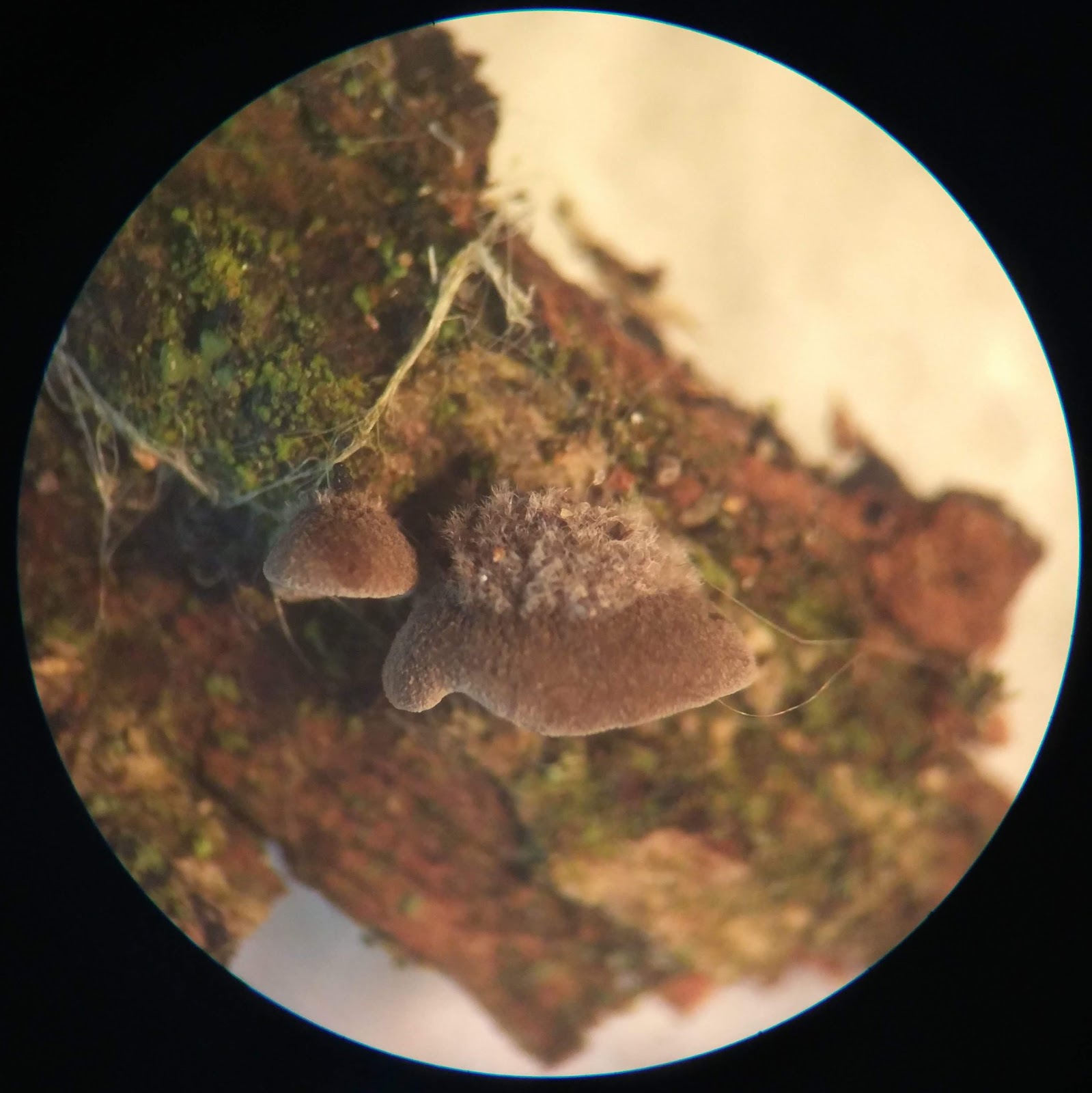
On a wet rotting branch nearby I spied some small grey 'crepidotoid' gilled mushrooms, growing in a small tiered cluster. Each one was no more than a few millimetres across. Looking closely, they all had this tufted hairy covering, towards the centre of the cap. In the older specimens the hairy part of the cap appeared darker, giving the fruit bodies a two-tone appearance.


I think these must be Hairy Oysterling Resupinatus trichotis. Quite an enigmatic little thing, when you get a good look at it.


On a large rotting log, at the edge of a sunken lane, we found a big patch of Tripe Fungus Auricularia mesenterica. A nice find as I've never seen this species before.


And another jelly fungus on Hazel...


Not sure where to start with that one.

For the record
Date: 27/07/2019
Location: Bonhurst Farm, Surrey

Records to be submitted via WWFRG

Thursday, 20 June 2019

Lundy Fungi

Fungus observations from LUNDY


Had a nice trip across the water to Lundy earlier this week. Splashed out on a copy of 'Lundy Fungi' by John Hedger & David George (published by The Lundy Field Society) which turned out to be money well spent as it's a really nice, informative photographic guide to the species you're likely to encounter in the island's different habitats.

Much of Lundy is designated as a SSSI and, as I was just there as a day-tripper, I'd made no effort to obtain the necessary permissions to collect specimens for identification purposes. What follows is therefore an account of what I was able to observe of the island's fungi in situ.

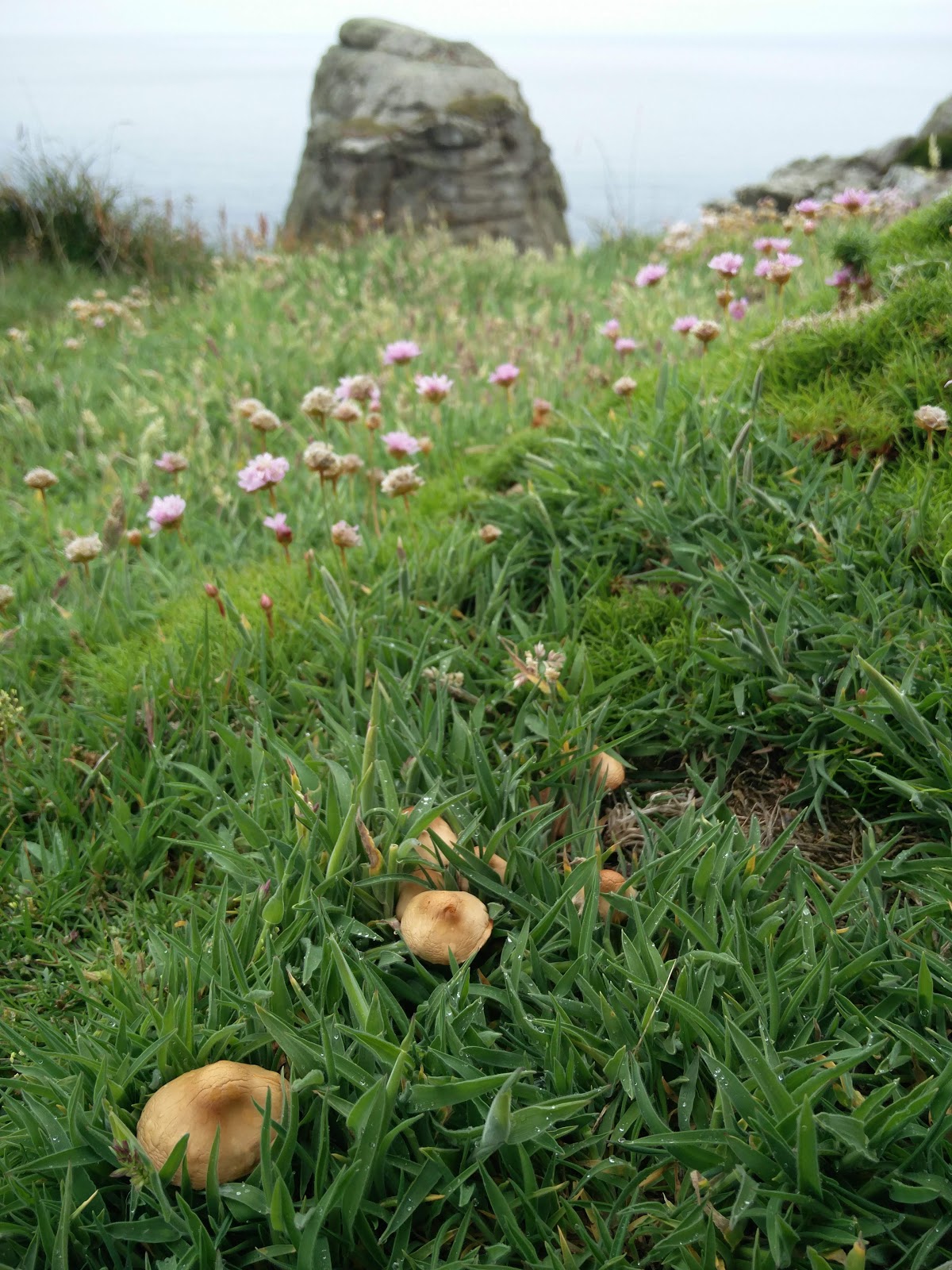
I'm reasonably certain the mushrooms in the photo above are Fairy Ring Champignon Marasmius oreades, with that pronounced umbo. They were growing in the short grass on the cliff-top, near Devil's Slide.

The grazed areas we passed through on our walk up the western coast were dotted with piles of dung, hosting some impressive fungal displays.

I think these pair are Egghead Mottlegill Panaeolus semiovatus...


The stems were dusted with black spores...


... and this younger specimen shows the ring around the stem nicely.


These, I'm sure, are something different. But I would not be confident in naming them.


I noticed that the young fruit body emerging towards the centre of this photograph had a distinctive reddish-brown colour to it.
 

UPDATE 20/06/2019 - advice from Mandy Dee, Lundy Fungus Recorder, over on the British Mycological Society Facebook page, is that this is Psilocybe coprophila

I suspect this yellow fellow is something different again. But hard to say without a closer look.


UPDATE 20/06/2019 - advice from Mandy Dee is that this is Stropharia semiglobata. A species which the Lundy Fungi book describes as very common on Lundy.

On the heath, near Jenny's Cove, I spotted quite a few of these guys. I think they're Heath Navel Lichenomphalia umbellifera.


And the Golden Hair Lichen Teloschistes flavicans at the North End was pretty special.


Heading back towards civilisation, on Hangman's Hill, I was happy to come across Campion Anther Smut Microbotryum lychnidis-dioicae again (after my recent encounter close to home).


 And there was the ubiquitous bramble rust...


I did allow myself to collect one of the bramble leaves, so I could get a closer look.

With reference to Ellis & Ellis's 'Microfungi on Land Plants', I think my collection is Phragmidium violaceum, with its mostly 3-septate teliospores. A new one for me.


For the record
Date: 18/06/2019
Location: Lundy, Devon

Thursday, 13 June 2019

Campion Anther Smut


I've been taking a closer look at the Red Campion Silene dioica on my route to work, ever since I heard about Campion Anther Smut Microbotryum violaceum sensu lato (from @fungifrolics). Finally found some! On Sussex Wildlife Trust's Woods Mill nature reserve, near the willow tunnel.

What mycological madness is this, to try and wrap my head around?! This is a fungus which, having infected a host plant, proceeds to take over the plant's sexual parts for its own reproductive purposes. In the place of anthers, which would have borne pollen, the fungus causes the plant to produce a mass of dark brown spores.


An illustrated life cycle of Microbotryum can be found in Schäfer et al (2010). Fascinating stuff!

There is also a very accessible introduction to the Microbotryum violaeum species complex on A. J. Silverside's website, here. And it seems that modern molecular studies have not yet completely succeeded in resolving the complex into a group of satisfactorily delimited species (Vánky, 2004).

I had read that the spores of Microbotryum are ornamented with a reticulate ('net-like') mesh. This prompted me to spend a few hours playing with the microscope.


Here are a couple of spores from my Woods Mill collection. Both images were taken at 1000x magnification under oil immersion & manually focus-stacked using Helicon Focus.
  • Left: on my trusty, somewhat ancient, SP03 
  • Right: on the older-model SP100 I recently acquired. 
Took me all evening, but I was happy to get that relatively crisp right-hand image from my 'new' microscope. It was interesting to compare with these SEM images in Denchev et al (2009).

Figures 3-4 from Denchev et al (2009). Spores of Microbotryum lychnidis-dioicae (Liro) G. Deml & Oberw. on Silene latifolia in SEM. Bars 3 = 1 μm, 4 = 2 μm. Image © Denchev et al 2009.

Having been thoroughly distracted by this fungus's biology and rather attractive spores, all that remains is to put a species name to it. Spooner & Legon (2006), in their list of species in the Microbotryum violaceum complex which can be recognised in Britain, have M. lychnidis-dioicae occurring in anthers of Silene dioica. Whereas Vánky (2004), advocates using the name M. violaceum sensu lato, pending further taxonomic work. So there we go. Take your pick.

There are a whole bunch of different Microbotryum species, parasitising different plants. I think one of the most modern comprehensive treatments must by Vánky (1998) but there is also a nice list with pictures! in the recent 'Smut and Allied Fungi of Wales' (Woods et al, 2018). Want to find them all now!


References

Denchev, C. M., Giraud, T. & Hood, M. E. (2009). Three new species of anthericolous smut fungi on Caryophyllaceae. Mycologia Balcanica. 6: 79-84. URL: http://max2.ese.u-psud.fr/publications/Denchev_2009.pdf

Schäfer, A., Kemler, M., Bauer, R. & Begerow, D. (2010). The illustrated life cycle of Microbotryum on the host plant Silene latifolia. Botany. 88. 875-885. 10.1139/B10-061. URL: https://www.researchgate.net/publication/235119641_The_illustrated_life_cycle_of_Microbotryum_on_the_host_plant_Silene_latifolia 

Silverside, A.J. Microbotryum violaceum. URL: http://www.bioref.lastdragon.org/Pucciniomycotina/Microbotryum_violaceum.html (accessed June 2019)

Spooner, B.M. & Legon, N.W. (2006). Additions and amendments to the list of British smut fungi. Mycologist. 20(3). pp. 90-96. URL: https://www.sciencedirect.com/science/article/pii/S0269915X06000322

Vánky, K, (1998), The genus Microbotryum (smut fungi). Mycotaxon 67: 33-60.

Vánky, K., (2004), Anther smuts of Caryophyllaceae. Taxonomy, nomenclature, problems in species delimitation. Mycologia Balcanica 1: 189–191. URL: https://zenodo.org/record/2546766#.XOOzcdh7m70 

Woods, R.G., Chater, A.O., Smith, P.A., Stringer, R.N. & Evans, D.A. (2018). Smut and Allied Fungi of Wales: A Guide, Red Data List and Census Catalogue. Aberystwyth. URL: https://eprints.soton.ac.uk/425372/1/smut_RDL.pdf

For the record
Date: 20/05/2019
Location: Woods Mill, West Sussex
Grid ref: TQ217137