Thursday, 12 October 2017

Worth Park fungus foray - Sunday 1 October


Fungus season was set to 'GO' in Sussex as we entered October. So much so, it's been a struggle to keep up with everything I've seen and attempted to identify. But I thought it would be worth sharing a few highlights from the foray at Worth Park, Crawley, with Nick Aplin of Sussex Fungus Group. After a busy couple of weeks, I'm now a bit hazy on which photos match which species listed in my notebook, so if you notice I've got some wrong, please tell me!

We met at Ridley's Court on Milton Mount Avenue. As soon as we stepped outside, we found mushrooms popping up in the wood-chipped flowerbeds and in the grass.

Nick identified this one as Redlead Roundhead Leratiomyces ceres.


Nearby, a fresh crop of Poisonpie Hebeloma crustuliniforme; looking a bit more impressive than the specimen we found at Tilgate Park the day before. You can just about see the moisture beading on the gill edges, and turning to brown specks, which is a key feature of this species.


At the end of Milton Avenue, there's a loop in the road which encloses a small green space, with three large cedar, cypress and redwood trees.

Under the cedar, we found an impressive patch of white mushrooms: White Dapperling Leucoagaricus leucothites.


Its slender stem features a white ring, which you can see here. The white gills are also a key feature.


Here we also found a Lepiota which I've been trying to identify. Nick Aplin's been in touch to say he's inclined to accept my tentative ID of Lepiota ochraceofulva, which is encouraging.

I've come across a few different Inkcap Coprinus-type mushrooms this year (including several which popped up in my village) but this was a new one on me: Hare's-foot Inkcap Coprinopsis lagopus.

We saw the mature fruit bodies first: stretching out their caps.


But nestled in the grass were also a few young fruit bodies, showing the furry-looking cap surface which gives this species its name.


Moving into the park, we came across a very fruitful area with a number of different Amanita, Lactarius and Russula species. This one, although rather battered, was interesting to see: Orange Grisette Amanita crocea. (I've come across Tawny Grisette A. fulva before, but never this species.)


Also in the parkland, we came across this 'mystery mushroom' which Nick Aplin later identified as Dark Fieldcap Agrocybe erebia.


There were some nice mushrooms in the short turf of Worth Park: indicators of old, species-rich grassland.

I think Nick identified these as Golden Waxcap Hygrocybe chlorophana.


And the pretty-in-pink Pink Domecap Rugosomyces carneus.


I think Nick said this was the Crazed Cap Dermoloma cuneifolium. Another species of unimproved grassland.


Moving on from there, we found a patch of Slimy Waxcap Hygrocybe irrigata.


This species was so slimy I had a hard job putting it down! The mucus on the cap kept sticking it to my hand.


We found Inocybe geophylla again, a species we'd come across at Tilgate Park the day before. This time we found the white version, White Fibrecap Inocybe geophylla var. geophylla growing under Lime.


And another grassland species which seemed like it should be fairly memorable, with it's slight build and slender stem: Butter Waxcap Hygrocybe ceracea.


While the group were busy looking at some brown Cortinarius species, one of our party ventured off into the shrubbery...


Upon his return, he mentioned quietly to me that he'd found something. And that something turned out to be a decent-sized patch of "EARTHSTARS!" I exclaimed, and danced a little jig.


Collared Earthstar Geastrum triplex.


This is the first time I've come across earthstars on my fungus forays, so a real treat to see.

Back at the Ridley's Court Visitor Centre, Nick had set up the microscope, ready to take a look at the microscopic features of our collections. I think this was a cystidium of one of the Inocybes up on screen, looking like... well... whatever it is an Inocybe cystidium looks like.


I was very taken with these: the cystidia of a Conecap Conocybe species.


They reminded me of the playing pieces from my old Cluedo set.
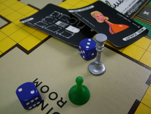
Image by Steve Berry | CC BY-NC-SA 2.0
Image by Steve Berry (CC BY-NC-SA 2.0)

Here are our collections from the foray laid out, so we could have a good look at their features and confirm identifications. A full species list has been circulated on the Sussex Fungi Yahoo! group (free to join if you're interested in recording fungi in Sussex).


For the record
Date: 1/10/2017
Location: Worth Park
Grid reference: TQ2938

All records to be submitted by Nick Aplin on behalf of Sussex Fungus Group

Monday, 9 October 2017

Mycena pearsoniana: Not a first for Suffolk

We didn't have ideal conditions for our fungus forays around Flatford Mill at the weekend. An extended period with little rain had left the ground very dry and some of Geoffrey Kibby's usual hunting grounds were almost entirely devoid of mushrooms.

But you can see too many mushrooms, and the advantage of sub-optimal conditions is that it gives you the chance to examine what fungi you do find in detail. It can also cause you to pay attention to things you might have otherwise passed by.

And so it was with this pinky-purplish mushroom, not much more than 1cm across, which was spotted growing in tussocky, scrubby grass...


... right about here (TM079330), on the thin strip of grassland between the footpath and the eastern side of the pond:


As well as its distinctive colour, this mushroom had a couple of other distinctive features: a stripey ('striate') margin to the cap; and decurrent gills which had clearly once run a little way down the stem, although now broken away.


Geoffrey provisionally ID'd it as Mycena pearsoniana, a species he has only come across a handful of times in 53 years of foraying. Which had the effect of making this small, rather dull-looking mushroom suddenly VERY EXCITING.

Once back in the classroom, we compared our find with the description of M. pearsoniana on the excellent 'Mycenas of Northern Europe' site, here. The macro-features all seemed to fit.

With respect to micro-features, the description talks about cheilocystidia "forming a sterile band" which I think is what I was looking at here, on the gill edge.


The cheilocystidia look like this, which I think puts them in the region of 'subfusiform' (albeit some of them look like they're verging on 'utriform').


The spores are fiendishly difficult to see, and impossible to photograph, but I think I've succeeded in measuring a few and they fit within the range for M. pearsoniana: 6 - 9 microns. There is no colour change in Melzer's Reagent.

All these micro-features seem like a decent match for M. pearsoniana. Hurrah!

Based on an initial look at records on the FRDBI database, we thought this could be a first record for Suffolk. However, on a second look I see there is one record for East Suffolk (VC: 25), collected by A. Henrici and identified by S. E. Wells from 2009. Still, a real rarity!

After my casual mention of a possible first for Suffolk on my last blog (here) I was contacted by Neil Mahler, the County Fungus Recorder, requesting details. In an exchange of emails, Neil very helpfully explained some of the background to fungus recording in Suffolk. Two mycologists called Martin & Pam Ellis produced a county checklist in 1988 (available on the Suffolk Biodiversity Information Service website, here). The checklist contains three records of M. pearsoniana: one found by the Ellis's from nearby Dunwich Forest; one from East Bergholt near Flatford by "A. J. Parker-Rhodes and his students"; and one from Staverton Thicks on a BMS foray with the aforementioned mycologists all present. The East Bergholt collection was recorded at Fishpond Wood, about 2 km from where we found it!

I wonder if there is a typo in the rendition of "A. J. Parker-Rhodes" name in the back of the Suffolk checklist, as the introduction talks about a Dr A. F. Parker-Rhodes who "organised fungus study weeks at the Flatford Mill Field Study Centre, foraying extensively in the surrounding area". Wikipedia relates that Arthur Frederick Parker-Rhodes (21 November 1914 – 2 March 1987) tutored a course on fungi at Flatford Mill for nearly 30 years.

Unfortunately the Suffolk checklist doesn't include dates for the records, but it must have been more than thirty years ago that Dr A. F. Parker-Rhodes was tramping around Flatford Mill with his students in tow, and found M. pearsoniana.

So, not a first for Suffolk. But a rather nice reminder that we walk in the footsteps of mycologists who have gone before us. Sometimes quite literally.

With very many thanks to Geoffrey Kibby for identifying this little mycena, and to Neil Mahler for putting this observation into context.

Specimen retained (Ref. CMB00002) and record to be submitted to the FRDBI in due course.

Record entered into FRDBI 07/09/2018

Saturday, 7 October 2017

Flatford Fungi

It's rather dry here in East Bergholt, Suffolk, but we still managed to find some cracking fungi today on Geoffrey Kibby's 'Identifying Fungi' course here at Flatford Mill.

With a fungi foray this morning and a fungi foray this afternoon, interspersed with classroom sessions on mushrooms and the basics of field mycology, topped off with an introduction to fungal microscopy, it's been a pretty fungus-filled day. I won't try and relay everything I've learned, because it's been SO MUCH! But I thought I'd share a few of the more characterful fungi that Geoffrey introduced us to, while they're fresh in my mind.

In one of the lawns near the field centre, Parrot Waxcap Gliophorus psittacinus; in shades of orange and green. Just gorgeous!


And nearby, in an area of dense, rank vegetation, Geoffrey spotted this: Blue Roundhead Stropharia caerulea.


"But that just looks a bit brown and boring", I hear you cry. Yeah, well, check this out:


The young fruiting bodies are BLUE, with pale grey gills. What a transformation.

Growing on a pile of felled elm logs, where Geoffrey has seen it before in previous years, we found some lovely Wrinkled Peach Rhodotus palmatus.


I've been wanting to see this species for a while and that young fruit body on the left didn't disappoint, with it's super-wrinkly cap.

Another species of rank grassland which we came across a few times today was Stubble Rosegill Volvopluteus (=Volvariella) gloiocephalus.


This species is notable for having a 'volva' – a bag-shaped structure at the base of the stem. A feature I'm used to seeing on Amanitas.


The volva is pretty thin and fragile though, so not always very easy to see. Makes you realise how closely you have to observe fungi in the field, to get a confident identification.

This afternoon we came across an impressive patch of Stinkhorn Phallus impudicus, growing in woodland.


Here's Geoffrey talking us through how the young fruit bodies develop, inside these egg-shaped structures.


Look at it!


On the way back we came across the Slender Parasol Macrolepiota mastoidea growing among the nettles in the corner of one of the sheep fields.


The stem surface, which is covered in small, closely-spaced brown scales, is one of the features which separates this from similar species like the Parasol Macrolepiota procera, which I've seen before (I think).


I kind of wish that whichever modern mycologists came up with common names for these species had some of the brazen literalism of 18th and 19th century fungal taxonomists who basically called these last two fungi (if you translate the latin): "Shamelessly Penis-shaped Fungus" and "Breast-shaped Mushroom". But I suppose that was in the days when ladies were not involved in mycology, and one has to behave with more decorum these days.

I might have also found a first for Suffolk, but more of that another day.


Records entered into FRDBI 07/09/2018

Friday, 6 October 2017

Friday night at Flatford Mill


I'm at Flatford Mill in Suffolk this weekend for a course on 'Identifying Fungi' with Geoffrey Kibby – author of many fungus identification guides, including the new 'Mushrooms and Toadstools of Britain & Europe'. So I'm hoping for 48 hours of fascinating fungus facts, from here on in.

After some basic introductions, Geoffrey began with a talk this evening on non-native and invasive fungi. Not a subject I've ever given much thought; but it was REALLY INTERESTING..


The basic premise of Geoffrey's talk was that the modern horticultural practice of mulching flowerbeds and shrubberies with woodchip, combined with a generally warming climate, is creating ideal conditions for non-native and potentially invasive fungus species to grow and spread.

It seems that very little is known about how the process of creating, transporting and spreading woodchip around is affecting species and habitats. And the risks, e.g. in enabling the spread of pathogens and aggressive non-native fungi, have not really been examined.

This struck a chord with me as I've been guilty of dumping a load of woodchip mulch on our garden at home, to keep down the weeds. Even when I noticed masses of not-previously-seen fruit bodies popping up in all the flowerbeds (here), I never really gave a second thought to what broader impact this might be having. It's easy to get fixated on looking for mushrooms and other fruiting bodies, and forget that fungi are ever-present, doing their thing, under the ground. (As an aside, I was listening to Lynne Boddy on The Life Scientific on the way here, talking about fungal communities, and she made this point very well: fungi live complicated lives which, to a very great extent, we don't yet really understand.)

Geoffrey proceeded to profile a number of woodchip-loving mushrooms which are spreading through Britain. These include:

  • Mulch Fieldcap Agrocybe putaminum
  • Redlead Roundhead Leratiomyces (=Stropharia) aurantiaca 
  • Blueleg Brownie Psilocybe cyanescens
  • Cucumber Cap Macrocystidia cucumis
  • Wrinkled Fieldcap Agrocybe rivulosa
  • Warty Cavalier Melanoleuca verrucipes

Classically alien-looking species like Devil's Fingers Clathrus archeri, Starfish Fungus Aseroe rubra and Red Cage Clathrus ruber also got a mention.

And it's not just the distribution of species which is changing. Geoffrey referenced a unique longditudinal study of fungal fruiting periods, based on data collected by fungus recorder E.G. Gange, which showed the UK fungus fruiting season is now around double the length of what it was in the 1950s; and numerous species are fruiting twice over the course of one year. There's a neat summary of this study on nature.com, here.

But it's not just us humans spreading fungi around. Species which were thought to be extinct in Britain, like the Pepperpot Myriostoma coliforme, and continental species like the Bearded Amanita Amanita ovoidea are present in a few sites in Britain and could spread, if climatic conditions become more favourable for them. Perhaps Caesar's Mushroom Amanita caesarea may make it to these shores. That's one mushroom it's worth keeping an eye out for! (You can find a description here).

Environmental change brings consequences, and Geoffrey finished by highlighting a few such consequences, from increases in pathogens and allergens, through to broader ecological impacts. It's clear there is much research that needs doing. And we'll need more mycologists in order to do it. 


I did warn Geoffrey I might be writing a blog, so I hope he will forgive me for stealing / sharing his content. Time to get some kip.

Monday, 2 October 2017

Trying to ID Lepiotas

I wouldn't recommend reading this post, unless you fancy helping me ID some Lepiotas collected from Tilgate Park and Worth Park at the weekend.

Lepiota 1


Location: Among grass by the entrance to the Walled Garden. Tilgate Park.
Odour: No particular odour detected.
Cap: Centre pale brown, cracking into concentrical, patchy scales, revealing white underneath.
Stem: White, silky.
Spore print: Creamy-white.

Cap without greenish colours
Cap and/or stem with brown to orange colours


Pileipellis... This is a thin 'scalp' section off the top of the cap. Can't really figure out what I'm looking at here. Is it a hymeniderm? It doesn't really look like the illustration in Funga Nordica (26C).

Can't remember if this was 400x or 1000x magnification.
Let's say "yes", pileipellis is a hymeniderm.

Spores: Dextrinoid. 7-8 microns long and approx 3 microns wide. Kind of bullet shaped with a spur on one corner of the flat end.

1000x magnification. Mounted in water.

1000x magnification. In Melzers (showing dextrinoid reaction).

Spores match L. cristata (543E)


Is this a bunch of cheilocystidia? If so, they're kind of maraca-shaped ('clavate').

1000x magnification.
I think we can say this one's definitely L. cristata. Assuming I got the bit about the pileipellis being a 'hymeniderm' right?

Lepiota 2


Lower stem under the stereomicroscope, showing patches of cap colour.


Location: In woodland, under Alder. Tilgate Park.
Odour: No particular odour detected.
Cap: Centre dark brown. Covered in brown pointed scales, with brown (you could possibly describe it as bronze, at a push?) colour extending all the way to the margin.
Stem: Beige-brown at the apex. Then brown further down, with patches of cap colour.
Spore print: Creamy-white.

Cap without greenish colours
Cap and/or stem with brown to orange brown colours 

Pileipellis... This is a thin 'scalp' section off the top of the cap. Again, don't really get what I'm looking at here. I'm gonna go with...

Pileipellis of elongate cells


I think this was at 1000x magnification.

Spores: Dextrinoid. 9 - 9.5 microns long and 3-4 microns wide. Spurred, with a more pronounced bulge off to one side, compared to collection above. Looks more like illustration for L. grangei in Funga Nordica (541E), although not as pointed at the tip.


Spores spurred.

Cap warm orange-brown (but doesn't appear to discolour yellowish orange on handling)

This takes me to a description of L. boudieri.

Some elements appear to match:
  • Pileipellis elements are septate, with clamps (yes, I think so, see below)
  • Spore shape and size is a reasonable match
  • BUT cap description seems a bit off; and stem in this collection is not 'widening towards base'


I think I got a look at some cystidia here (hard to see, as they didn't take up the stain) - cylindrical to clavate?


Not too confident with this one. I think I've probably gone wrong because the collection doesn't look like photos of L. boudieri.

Lepiota 3 

Pretty sure this one's L. cristata again, as Nick observed smell of 'coal gas'.


Location: Pathside. Tilgate Park.
Odour: 'Coal gas'

Spores 6-7 microns long and approx 3 microns wide. Spurred.


Pileipellis


Cystidia?

Basidium?

Lepiota 4 


Location: Worth Park
Odour: Yes. Just smells mushroomy to me.
Cap: Centre brown, turning beige towards the edges (not white). Some concentric cracking around the centre, but not as patchy as in (1) and (3) above.
Stem: Cream at the apex to brown lower down, with girdles of (beige) cap colour around the lower half of the stem.
Spore print: Creamy-white.

Cap without greenish colours
Cap and/or stem with brown to orange colours 

Pileipellis


A hymeniderm?

Spores Dextrinoid. 7 microns long and 3-4 microns wide. Ellipsoid.


Spores ellipsoid

I think this is an image of cheilocystidia, stained with PlaqSearch.


Cheilocystidia present

This takes me to L. ochraceofulva.
  • Spore shape and size matches.
  • Cheilcystidia narrowly clavate.
  • A few scattered girdles of cap colour at the base. 
  • Gills remote from stem.


However, other features seem like not such a good match:
  • Not seeing shades of orange / yellow
  • Looks to me like it turns brown with handling (not orange)
  • There is a smell to it, but I'm not getting 'orange chocolate' or 'unpleasant'
I also can't see any previous records from West Sussex for L. ochraceofulva, which makes me suspicious I've gone wrong somewhere!

If you've got this far - thank you! - any advice gratefully received.