Saturday, 23 September 2017

Selwyns Wood


I joined the Sussex Fungus Group foray at Selwyns Wood last weekend. This is a Sussex Wildlife Trust nature reserve which I don't think I've ever been to before, so I thought I'd better rectify that. We were met by Trevor Hince, the Volunteer Reserve Manager, who had very kindly offered to take us round.

The first part of our route took us through mixed woodland, where we found The Deceiver Laccaria laccata popping up in various places...


... Along with it's more bohemian cousin, the Amethyst Deceiver Laccaria amethystina.


I was thrilled to see the Parasitic Bolete Mushroom Pseudoboletus parasiticus: a species I've not come across before.


It's easy to identify because it grows from the base of Common Earthball Scleroderma citrinum.


We were under instructions from the Sussex Wildlife Trust ecologist, Graeme Lyons, to look out for Rutstroemia echinophila, a brown disc-shaped ascomycete that grows on last year's Sweet Chestnut husks. And here it is:


I think Graeme said there was a cool vernacular name for it, like Hairy Nut Cup, or something. But I've forgotten it.

Nick Aplin also found this, which he identified as the rather scarce Plicatura crispa.


When we spotted this faded yellow Russula, I thought it would be a good opportunity to try out my new iron sulphate crystal (which has a pleasing resemblance to kryptonite).


The genus Russula (the Brittlegills) contains many very similar-looking species and you have to observe them very closely to be confident of an identification. One of the features you can observe, which is well documented in Geoffrey Kibby's new 'Mushrooms and Toadstools of Britain & Europe Volume 1', is the reaction of the fruit body to chemicals such as FeSO4: iron sulphate. Some Russula fruitbodies will display a colourful chemical reaction when rubbed with an iron sulphate crystal, turning shades of green, yellow, orange or pink.

I rubbed the stem with my crystal, as if it were a magic lamp, and wished for a spectacular reaction.

Slowly – in a not-very-impressive way, if I'm honest – the stem turned a sort of pinky-brown color, where I'd rubbed it.


Others in the group identified this Russula as the Ochre Brittlegill Russula ochroleuca; and we moved on.


I wish now I'd taken a specimen, as it turns out the fruitbody might have done something more spectacular if I'd applied KOH to the base of the stem: i.e. turn BRIGHT RED!


Reproduced from 'Mushrooms and Toadstools of Britain & Europe Vol. 1' by Geoffrey Kibby


I should have also given it a sniff, as there's another similar-looking species which smells of pelargoniums (geraniums). It seems I wasn't really at the top of my fungal observation game last weekend and, after a busy week, I've forgotten some of the details of what we saw / sniffed. Really must write things down!

Rounding the corner, we emerged in an area of heathland, which was a nice surprise.


I didn't note much here fungus-wise. But we did find this Brown Rollrim Paxillus involutus. You can see where it gets its vernacular name.


From there we moved into an area of Sweet Chestnut coppice which proved to be the most fruitful habitat of our foray.

False Deathcap Amanita citrina popped up in a few places, with its odour of raw potatoes.


There were a few birch trees around, at the edge of the Sweet Chestnut coppice, and we made this Brown Birch Bolete Leccinum scabrum. The dark scales on the stem are a key feature of that species.


The stem flesh showed no hint of blue when cut.


All through the Sweet Chestnut coppice there were little red Russulas popping up through the moss that covered the old coppice stools. I sadly failed to get a photo of these. I'm also rather disappointed I didn't get a photo of Janet's face when she tasted one

Growing amongst the leaflitter, we came across this rather non-descript-looking mushroom which Nick persuaded me to take as a specimen, to see if I could get any sense out of it down the microscope. I forgot to take a photo in situ, so this is what it looks like after it's been sitting in my fridge for a week.


It's kept remarkably well. Up close, the wooly ('floccose') stem looks quite distinctive. The cap, too, looks like it was covered in fine, silky (?) hairs.

Under a hand lens or stereomicroscope, you can see the mouse-brown gills have a white edge to them.


I think Nick gave me a clue about what genus we're in here, but I didn't write it down (what was I doing?!). So let's see where MycoKey takes us...

Psathyrella

OK, so, the first question in the key to the Psathyrella in Funga Nordica asks if the spores are > 9μm or < 9 μm. I haven't got a spore print, because the mushroom has been sitting in a box in my fridge for a week. So I'll have to make do with measuring the spores from a gill squash.

I've measured ten spores and I make them all about 9 μm. This is not a good start.

Not wanting to admit defeat at such an early stage, I've decided that - if there's anything in it - the spores are probably a fraction under 9 μm; rather than over. This takes me to a question on whether 'pleurocystidia' (cells on the face of the gills) are present or absent. Yikes! I think this means I'll have to try and look at a cross section through the gill: a gill trama squash. Tricky...

I'm pretty sure I can see pleurocystidia on the face of the gill, but they're rather difficult to see with all those spores washing around.

 
I thought I'd try staining a gill trama squash, and see if that makes the pleurocystidia any easier to see.


YES! This is a cross section of one of the gills at relatively low magnification (100x). I've stained it with PlaqSearch, which was recommended to me as a good, safe, general purpose stain, and then washed it back through with water. On the left hand face you can see lots of vaguely skittle-shaped cells which actually haven't taken up the stain particularly strongly. I think these are pleurocystidia.

Here they are at 400x magnification. Is that 'incrusting' that I can see on them?


Anyway, back to the key.

Now I've got to see if the gill edge and cystidia are "covered in drops staining green in a solution of ammonia." I did this the other day. I can do this!


It hasn't stained green.

This takes me to the following entry in the key:
  • Veil absent; pleurocystidia with crystals
  • Veil present; pleurocystidia with or without crystals
Hmm. This is where I wish I'd more closely observed the mushroom when it was fresh. When you look closely at the edge of the cap, there is something which looks like it could be the remains of a veil.


And crystals. Hmm... that must be what I observed above: pleurocystidia with crystals. I'll go with the second option: "Veil present; pleurocystidia with or without crystals."

I don't think this mushroom smells "sweetish"; so now I have to look at the shape of the pleurocystidia.


Hmm. This is really tricky. I'm going to go to key J: "Pleurocystidia lageniform to conical, upper part often acute."

This takes me to Psathyrella cotonea, but – looking on vielepilze.de – that doesn't seem right to me. 

*many hours pass, going backwards and forwards through Psathyrella keys...*


I tried mounting the spores in KOH to see if that would help guide me towards an identification. I discovered that the spores turn darker brown in KOH; but it didn't really help!

 
I've now trawled through various other possibilities (P. fibrillosa, P. impexa, the list goes on!) and I think it's time to admit I'm stuck with this one. Nick Aplin also took a specimen, so here's hoping he managed to get to an ID.


As we headed back towards the car park, we had a sudden rush of gorgeous Cortinarius species. I generally steer clear of attempting to identify Cortinarius (Tuula Niskanen's talk at Kew last year rather scared me off) so it was nice to be introduced to these beauties.

Saffron Webcap Cortinarius croceus


Frosty Webcap Cortinarius hemitrichus


And finally, the Surprise Webcap Cortinarius semisanguineus.


 Why is it a surprise? You may well ask. Here is Nick Aplin, about to do the big reveal...


 It's got bright red gills! Crazy!


The walk back to the car park produced a few more species for the list, including Fly Agaric Amanita muscaria popping up through the moss.


And another parasitic species: Silky Piggyback Asterophora parasitica, which grows on decaying Russulas.


Those are just a few of the highlights for me. Both Nick Aplin and Ted Tuddenham took specimens away to check under the microscope, so it's possible a few of the field identifications I've mentioned above may change.

If you'd like to see the full list from this Sussex Fungus Group foray, then sign up to the Sussex Fungus Group Yahoo! group.

For the record
Date: 16 September 2017
Location: Selwyn's Wood
Grid reference: TQ5520

Records will be submitted by Nick Aplin, on behalf of Sussex Fungus Group

Monday, 18 September 2017

Between a rock and a hard place


I passed this spectacular display of fungi on my way to work this morning, growing between a brick wall and the pavement.


I was just amazed to see this densely-packed mass of mushrooms, bursting through the tarmac.


The patch on the left were clearly a day or two older than the rest, in shades of brown and beige, while the younger fruit bodies were a bright, lustrous tan colour.


The youngest fruit bodies, where they had some shelter from the elements, looked like they had spiky little white skirts. What style!


I must be getting better at this mycology malarkey, because I think my initial guess at their identity a Psathyrella of some kind was correct. This strongly clustered growing habit is typical of the Clustered Brittlestem Psathyrella multipedata.

I had a look at one of the fruit bodies and it seemed a good match for P. multipedata.


If you look closely, you can see the cap is "faintly striate halfway towards the centre".

Funga Nordica describes the veil as "scattered fibrils near the margin": the spiky white skirt I'd seen in the photo above. There's a German website vielepilze.de which includes comprehensive information on the European Psathyrella; it describes the veil as "very volatile, mostly only visible in youngest stages, often absent" which also fits with what I've observed.


The young gills are pale brown and crowded.

The key to the Psathyrella in Funga Nordica (which you can access through MycoKey) says that the gill edge and cystidia are "covered with drops staining green in a solution of ammonia". I've got some ammonia, I bought a massive bottle of it at the hardware shop in the village. So I thought I'd give this a try.


I think this might be my MOST EXCITING GILL SQUASH yet. Not only is the gill edge covered in attractive bottle-neck-shaped ('lageniform') cystidia, but they went green! I quickly measured the spores as well, and made them about 7 microns.

I think I'm on pretty safe ground calling these mushrooms Clustered Brittlestem Psathyrella multipedata.

The habitat does seem a little odd though. Funga Nordica describes this species as occurring "on soil, usually attached to buried wood, on stumps, in forests or parks ..., [or] on calcareous or nutrient rich soil, in grass lawns and on sawdust". But Roger Phillips describes the habitat as "amongst grass in open deciduous woodland and on roadsides." – which fits better with where I found them.

For the record
Date: 18/09/2017
Location: Small Dole
Grid reference: TQ2113

Record entered into FRDBI 06/09/2018

Golden Spindles

Thought I'd go for a walk around the village to clear my head after this weekend's fungus identification marathon.

Couldn't resist snapping a photo of these little beauties, growing on someone's front garden.


I was going to call them Golden Spindles Clavulinopsis fusiformis; but then I noticed the Collins (illustrated) Fungi Guide includes another species, Yellow Club Clavulinopsis helvola, which is "very common; often confused with C. fusiformis but smaller and spores different".

These fruit bodies did seem quite small to me, so I wonder if what I've got here is C. helvola. Haven't had any luck getting a spore print, yet.

For the record
Date: 17/09/2017
Location: Small Dole
Grid reference: TQ2112

Sunday, 17 September 2017

Graffham Common Fungus Walk

Image © Katie Parker

I had my first experience of leading a fungus walk on Wednesday at Graffham Common – sharing what I've learned about fungi with some colleagues from the Sussex Wildlife Trust

Previously covered almost entirely in pine plantation and rhododendron, Sussex Wildlife Trust is in the process of restoring this site to wooded heath with an acid pond which is great for dragonflies. Ecological monitoring has found a fabulous diversity of rare plants and invertebrates here, and the site is also providing habitat for many notable birds – you can read more about this on Graeme Lyons' blog: The Lyons Den.

We started the walk on Gallows Hill. It says something about what an open landscape this must traditionally have been that this was once where criminals were once left to hang, where they could be seen from miles around.


View from Gallows Hill across Graffham Common.

I did warn my colleagues we'd probably find lots of mushrooms that I wouldn't be able to identify. But, fresh back from that fungal identification skills course in Barnsley, I decided I'd collect some specimens and have a shot at identifying them when I got home.

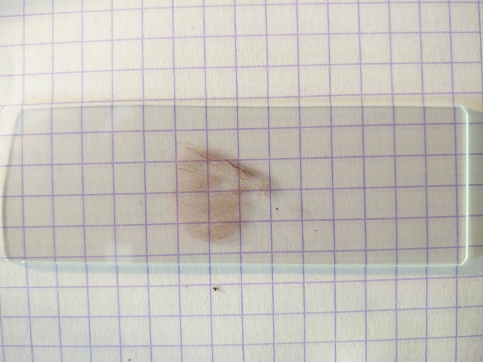
Here they are all laid out ready for step one: get a spore print!


The walk started off with a nice easy one to remember: Turkeytail Trametes versicolor, growing on the end of a rotting birch log.


On the same log we saw the smaller, orangey fruit bodies of Hairy Curtain Crust Stereum hirsutum. Always a crowd pleaser.

There were also earthballs Scleroderma sp. galore. Including this massive one.


I still haven't got my head around identifying earthballs to species. I they were probably all Common Earthball Scleroderma citrinum but not 100 % sure.

Then things got interesting!

We saw masses of these striking orangey-brown mushrooms trooping across the ground, amongst the pine litter.


The younger ones were very cute with their little button caps.


The mushrooms had streaky stems, with contrasting dark and pale fibres; as you can see here:


Here's what those gills look like in cross-section.


And they dropped a copious amount of white spores.


After a not-very-successful attempt at using the synoptic key to genus in MycoKey, followed by much flicking through my field guides, the penny finally dropped. They are a deceiver Laccaria sp. of some kind.

I narrowed it down to either The Deceiver Laccaria laccata, or the Scurfy Deceiver Laccaria proxima.

The Scurfy Deceiver Laccaria proxima is found on acidic soils, in coniferous woodland and on heaths. Which is exactly the sort of habitat we found these mushrooms in.

One article I read (on Michael Kuo's website, here) suggested you can tell the two apart by looking at the spores: "the spores of Laccaria laccata are round (or nearly so), and have fairly prominent spines; the spores of Laccaria proxima are mostly elliptical in outline, and have smaller, less prominent spines." I can do this! I thought. Until I tried to look at the spores: they are white and it's very difficult to see the spore ornamentation when they're mounted in water. I looked on the BMS website to see if they had any advice on what chemicals to use, to get a better look at the spores:


Phloxine B. Never heard of it!

But by this point I'd got all fired up about the prospect of seeing some spore ornamentation, so I thought I'd have a go at staining the spores with Cotton Blue and see where that gets me.


Hmm. Well. You can tell that the spores aren't smooth and, when you twiddle the focussing knob, you can just about make out some not-very-prominent spines. Plus they are mostly elliptical.

I think the balance of probabilities is tipping towards my collection being the Scurfy Deceiver Laccaria proxima.

It's worth pointing out that the key to Laccaria in Funga Nordica doesn't make much of the difference in the spores. The main features they use to separate the two are:
  • Whether the cap is "smooth to tomentose [covered in hairs]", or "scaly"; and
  • Whether the thin fibres on the stem are contrasting, or not.

Looking at the species descriptions in Funga Nordica, I'm pretty sure that what I've got here is the Scurfy Deceiver Laccaria proxima.

Next up, we saw quite a few of these yellowy-orange mushrooms, with a pronounced nubbin (or 'umbo') on top, growing on decaying conifer logs.


Image © Katie Parker
I couldn't see any trace of a ring around the stem.


And the gills were yellow; turning orange with age.


 These have produced a rusty orange spore print.


MycoKey worked well on this occasion, taking me to the rustgills Gymnopilus sp. This genus includes quite a few species which occur in Britain, many of which look quite similar.

From the Collins Fungi Guide by Buczacki et al.

Gymnopilus have cells on the edge of their gills, called 'cheilocystidia', which can have quite distinctive shapes. I think this is one, in the centre of this photograph:

1000x magnification; mounted in water and stained with Congo Red.

Given that these mushrooms were growing on conifer wood, and given the appearance of the cap and stem, I'm leaning towards this mushroom being Common Rustgill Gymnopilus penetrans. The shape of the cheilocystidia would fit with that, I think.

OK, next mushroom! This one was growing on an old burn site, on sandy soil.


It has a quite different shape to it, compared to the previous two mushrooms we'd looked at. I thought at first it might be a milkcap Lactarius sp. but it didn't produce any milk.


The spore print is white.


I think this takes us to the funnels Clitocybe sp.

Flicking through Roger Phillips' Mushrooms, there's one Clitocybe that looks like a good match for my mushroom: Clitocybe sinopica, which is found "in coniferous woods, especially in burnt areas". Although Roger Phillips notes that it's "uncommon".

A quick check on the old FRDBI database picks up only 132 records for this species in Britain, ever. But there are a handful from West Sussex, including a record from Ambersham Common in 1990 which is just up the road. So it's not entirely implausible. But if you're recording something that uncommon, you want to be sure that's definitely what it is.

Turns out one of the key features of Clitocybe sinopica is a strong 'farinaceous' (starchy) smell. Unfortunately I forgot to give it a good sniff out in the field and now three days later the mushrooms are rather dried out and don't smell of much. So I decided to seek some advice on the British Mycological Society Facebook page.

I got some advice from Ken Burgess, who suggested I start off by looking at the spores, to make sure I've definitely got a Clitocybe.

1000x magnification. Mounted in water.

I've got that problem of looking at white spores under white light again. But you can just about make out their elliptical shape. Inside the spores you can see little bubbles of varying sizes; these are oil droplets.

The next step is to measure the spores. For this I thought I'd stain them in Cotton Blue, to make them a bit easier to see.


I've previously calibrated my microscope, so I know that one minor unit on this scale is about 1 micron (1 thousandth of a millimetre). That makes the spores about 4 - 5 microns wide; and 6 - 7 microns long.

This then kicked off quite a lengthy discussion with Ken Burgess and Malcolm Storey, over on the BMS Facebook page, as it takes me into an area where there have been some recent taxonomic changes...

The spores are, arguably, a bit on the small side for Clitocybe sinopica and at some stage another (very similar) species was described: Clitocybe subsinopica, with smaller spores. So, based on spore size, my mushroom would seem a better fit for Clitocybe subsinopica. However, Malcolm Storey advised that, in a more recent round of taxonomic changes, these two species have now been lumped together again, under the name Clitocybe sinopica.

Indeed, Funga Nordica treats the two names as synonymous with each other:


Malcolm left me with a wonderful piece of advice, which fits rather nicely with the title of this blog:

"Only beginners are absolutely certain of their IDs. For the rest of us they're the best we can do with the books and papers available at the moment."
I was tempted to call this one Clitocybe sinopica and be done with it. But then Richard Shotbolt picked up that my spores have very strongly taken up the Cotton Blue stain. In mycology-speak, this makes them 'cyanophilous'. Richard pointed out that Kuyper, in his publication Flora Agaricina Neerlandica on the agarics and boleti occuring in the Netherlands, states that Clitocybe sinopica is "not cyanophilous". Which leaves the identity of this mushroom something of a mystery...


Right, well, if you can believe it, I've now been writing this blog for about three days. Time for a nice easy one:


Tawny Grisettes Amanita fulva were popping up all over the place. I remembered them from when I encountered them at Ambersham Common.

Then, growing on a sandy, moss-covered bank, next to the track, we saw these:


Here's what one of them looks like up close:


The spore print is a slightly-creamy white.


Flicking through my field guides, it looks to me like it could be something like the Heath Navel Lichenomphalia umbellifera. I'm not sure I've got any more microscopy in me right now, otherwise I'd check it.

Further down the track we encountered masses of what-I'm-assuming-is Sulphur Tuft Hypholoma fasciculare.


They were growing all along the edge of the track, at the base of the bank.


I've seen this species before, growing on rotting logs and wood piles. But I've never seen them growing like this. The Collins (illustrated) Fungi Guide says that Hypholoma do occasionally grow on "impoverished acidic soil".

I'm pretty sure they are Sulphur Tuft Hypholoma fasciculare...


There is another similar-looking species called Conifer Tuft Hypholoma capnoides which the first-nature website describes as having "pale grey gills with no hint of green". But the gills on my collection are definitely greenish.


We also found Earthfan Thelephora sp. growing in various places.


I think this is Common Earthfan Thelephora terrestris. But there is another species, the Carnation Earthfan Thelephora caryophyllea which looks similar. According to the Collins (illustrated) Fungi Guide, one has "warty to knobbly" spores, and the other has spiny spores.


I've got a spore print it's brown. So let's see if we can see any knobbly-ness.

They look pretty knobbly to me!


So let's call that one Common Earthfan Thelephora terrestris.

I'd like to say that's the end, but I did also find some other little brown jobs growing on the hard standing, which seemed to be made up of gravel and wood chippings, where I'd parked my car. There were quite a few, dotted around.


These are the kind of mushrooms I'd normally pretend I hadn't seen. But, armed now with some basic microscopy skills, I thought I'd give them a go. 

I think this two-tone coloration on the cap makes them 'hygrophanous', which just means the cap changes colour as it dries out.


The gills are fairly crowded.


The gill attachment is – I'd say – 'adnexed', i.e. narrowly attached to the stem. And, if you look closely, you can make out long streaks or grooves running down the stem, which is slightly bulbous at the bottom.


It has produced a fairly copious brown spore print.


After consulting with MycoKey, I think this may be one of the conecaps Conocybe sp. Another really difficult genus. I think I'll leave this one!

For the record
Date: 13 September 2017
Location: Graffham Common
Grid reference: SU9319

C. sinopica specimen retained (ref. CMB00001).

Records entered into FRDBI 06/09/2018