'Tis the season for having a go at botany: one fungal substrate at a time.
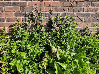
If you zoom right in on this sickly-looking Periwinkle (Vinca sp.) then you can just make out a hairy edge to the leaves...
I think this makes today's plant Greater Periwinkle Vinca major.
And if I've got that right, then I think those black pustules scattered generously across its leaves are one of the life stages of the rust fungus Puccinia vincae.
Under the stereo microscope (image below) you can see the hairy margin
of the leaf nicely and I think the orange specks are 'stage 0' of the
fungus's lifecycle: the spermogonia. The black specks are 'stage I': the
aecia.
One or two of the leaves are showing these gingery-coloured pustules, 'stage II': the uredinia.
That gingery-coloured stuff is actually a mass of urediniospores, pictured here at 100x magnification (image is of a cross-section through one of those pustules):
This is what the urediniospores look like at 40x magnification.
I really had to hunt for 'stage III', but eventually managed to track down a few solitary teliospores (one pictured below at 1000x magnification).
I read on the fungi.org.uk forum that the teliospores of Puccinia vincae look like peanuts, which gave me some motivation to fiddle around with oil immersion and a bit of focus-stacking. And I have to agree, it does look like a peanut.
***
That might have been the end of my study of Puccinia vincae but, what do we have here?
Some ashy-grey looking pustules (or, 'sporodochia'?):
I think this gives us the threefer! (If I am correct) what we have here is another fungus: Tuberculina sbrozzii, parasitising the rust fungus Puccinia vincae which is itself a biotrophic parasite on the Greater Periwinkle Vinca major.
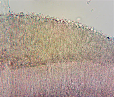
These ashy-grey sporodochia are structurally very different to the uredinia I was looking at earlier. Here is an image of one in cross-section at 100x magnification.
The outer surface is covered in little round conidia.
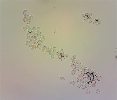
Here are a load I scooped out from the middle of a
sporodochium, at 400x magnification:
You can see that they are clear, or 'hyaline', and when I measured them they were about 10 microns in diameter - which is the right ballpark for Tuberculina sbrozzii.
For the record
Date: 8 April 2023
Private site in East Sussex
(Records will be entered into iRecord in due course.)


