Success! Although I struggled to get a decent photo in the low light.
The wet willows proved to be a pretty rich hunting ground for fungi.
This rather lovely purple thing is the Silverleaf Fungus Chondrostereum purpureum.
Here and there the branches were splashed with yellow.
Andy Overall wrote a very useful article on these 'Yellow Brain Fungi' in volume 18 (3) of Field Mycology [1]. There are two species which look very similar: Tremella aurantia and Tremella mesenterica and I've always been rather confused about how you separate the two.
Andy explaines that Tremella species are parasitic on other fungi or lichens, and the two species mentioned above are specific to different hosts. T. aurantia grows almost exclusively on Stereum hirsutum whereas T. mesenterica grows on resupinate (flat-growing) fungi from the genus Peniophora.
I've looked for a host fungus when I've found 'Yellow Brain' in the past and never been able to find any sign of it. In another paper on these species, which Andy references [2], Peter Roberts notes that T. aurantia "seems to enter the host fruit body whilst it is still developing, so that no external host normally remains" and T. mesenterica "appears to parasitise the mycelial hyphae rather than the fruit bodies of the host." So I don't feel so bad now about not being able to find the host.
Peter goes on to say that, "Often T. mesenterica can be found growing on the upper surface of a twig with the Peniophora producing fruit bodies on the underside".
I hunted around near where this 'Yellow Brain' was growing to see if I could find any sign of a host. I found this, a short distance away on the same branch.
Could this be a Peniophora? I'm rather intrigued by it, whatever it is, as the ashen-grey crust appears to be dotted with red blobs.
Both papers say that the two 'Yellow Brain' species can be separated with microscopy and there are some nice illustrations in Peter Roberts' article, so I feel like I should have a go.
 |
| Reproduced from Roberts, 1995. |
UPDATE 09/01/2018
Taking a tiny slither off the outside of that 'Yellow Brain' I was surprised to discover this tangle of threads (hyphae?) as well as the anticipated blobs (basidia? spores?).
 |
| 100x magnification. Mounted in water. |
I'm still getting the hang of using my new eyepiece camera so I was really pleased to find - amongst all that chaos - something which looked just like illustration 7 above: a basidium! The line down the middle shows it is 'septate'.
 |
| 400x magnification with zoom. |
I had a harder time finding the spores, but I think this is one:
 |
| Can't remember what magnification this was. |
I was struggling to see features clearly when mounted in water, so I thought I'd try them with a stain. I opted for PlaqSearch which I've been told is a safe, general-purpose stain.
Yup! That works!
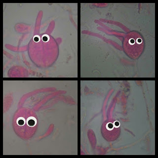
With everything stained this shocking pink colour, it was easier to spot those blobs with dangly bits on which look like a good match for T. mesenterica basidia.
These microscopic features seemed to have a lot of personality. But I felt like they were missing something... Googly eyes!
If you squint at this image, I think you can make out some clamped hyphae.
And I found these quite hard to find, but I think this image shows 'conidiophores' (whatever they are; bottom of illustration 8, above).
 |
| 400x magnification. Mounted in water; stained with Congo Red. |
For the record
Date: 6 January 2018
Location: Woods Mill
Grid reference: TQ219134
Record entered into FRDBI 07/09/2018
1. Overall, A. 2017, Tremella aurantia & T. mesenterica, two British 'Yellow Brain Fungi' compared, Field Mycology Vol. 18 (3), 2017
2. Roberts, P., 1995, British Tremella Species I: Tremella aurantia & T. mesenterica, Mycologist Vol. 9, Part 3 https://doi.org/10.1016/S0269-915X(09)80270-X


I have never seen the host near a yellow brain and wondered why. Thanks for highlighting Peter Roberts' explanation of that. The grey resupinates on the branch look like lichen to me. The red blobs could be their hymenial discs. I don't know any lichenologists down here. Is there one recording for the county?
ReplyDeleteThanks Ted. There isn't much active lichen recording happening in Sussex, as far as I'm aware. There is a county recorder - Simon Davey - but he's not able to get out in the field like he used to.
DeleteThis comment has been removed by the author.
ReplyDeleteAfter reviewing Nic's comment and looking at the picture again I can see that I had missed the pink patch with the red blob which Nic confirms as Peniophora like, and therefore a perfect association for T. mesenterica.
ReplyDeleteThanks Ted! I've had fun with this one. Just updated the blog with microscopy.
DeleteMy local grassland is surrounded by willows and I will taking a much closer at them look for fungi over the next few weeks. Also I'd like to say what an excellent blog! I am still very much a beginner and find your posts very informative. Thank you.
ReplyDelete